
Dr Cynthia K. Thompson – Innovation in Promoting the Recovery of Language after Stroke
Stroke can impair a person’s ability to communicate, resulting in a disorder known as aphasia. To facilitate recovery, scientists must understand how language is processed normally as well as how a stroke may impact the language system in the brain. Dr Cynthia K. Thompson, Ralph and Jean Sundin Professor of Communication Science and Professor of Neurology at Northwestern University, has been researching normal and disordered language for over thirty years. Her focus is on understanding and supporting the recovery of language processes when the brain has been damaged.
The Complexity of Language and the Brain
The uniquely human capacity for language resides in the most complex organ in the body, the brain. While significant insights have been gained in ascertaining the neurobiology of language, the inherent complexity of the brain, as well as language and linguistic processes, means much is still to be achieved. Damage to the brain can occur through a wide range of different diseases and injuries, and can have severe consequences for the language capabilities of an individual.
Aphasia Caused by Stroke Damage
Aphasia is a language disorder that results from brain damage. A major cause of aphasia is stroke, which affects over 15 million people across the world. Stroke is a life-threatening condition, in which blood supply to areas of the brain is cut off or significantly reduced. Reduced blood supply to regions of the brain that support language processing renders those regions unable to process language normally. It is estimated that around one-third of stroke survivors are left with aphasia, which affects the ability to understand and formulate language. This often has wide-reaching effects on the ability to communicate, which impacts social independence and the quality of life and increases the likelihood of social isolation.
Dr Cynthia K. Thompson at Northwestern University works to understand which areas of the brain are responsible for language processing – in particular, sentence processing – in healthy individuals to aid rehabilitation and recovery in those with deficits. By mapping the neural structures of sentence comprehension and production in healthy volunteers, her work is providing vital clues as to how networks may be rebuilt, potentially in different areas of the brain – a phenomenon known as ‘brain reorganisation’.
Many previous studies have used neuroimaging techniques to record the complexity of neural activity across multiple brain regions when language is processed. Dr Thompson and her colleagues published a review of previous studies observing the neural activity of people recovering from aphasia and healthy individuals. They found some important differences between the two groups. Healthy individuals typically displayed activity primarily in the left hemisphere of the brain when processing complex sentences whereas those recovering from aphasia were found to display greater activity in the right hemisphere. This observation supports Dr Thompson’s conviction that right hemisphere brain tissue may be recruited to support sentence processing in people with aphasia.
In further research, Dr Thompson has effectively demonstrated that language functions can indeed be regained with training. Dr Thompson also maintains that it isn’t just those who have recently suffered a stroke that can recover language skills. She has shown that patients can demonstrate significant brain reorganisation many years poststroke, allowing the return of at least some previous language skills and functioning. Dr Thompson explains her approach as follows: ‘If we can find the parts of the brain that are the most likely candidates to be recruited for recovery, then the next step is to push those parts of the language network of the brain using non-invasive neurostimulation.’

Neurocognitive Mechanisms Leading to Agrammatism
Dr Thompson and her team investigate the neurocognitive mechanisms that lead to a specific deficit known as agrammatism, a type of aphasia that affects grammatical aspects of language processing. Neurocognition refers to the study of cognitive functions that are closely linked to specific brain areas or pathways.
Dr Thompson and colleagues found that agrammatism is caused by damage to frontal and temporal lobe regions of the brain as well as the subcortical white matter fibres which link them (known as the dorsal pathway) in the left hemisphere, which is associated with language processing in the majority of humans. More specifically, these regions play an important role in using and understanding verbs and verb-argument structure within speech, which are crucial for the production and comprehension of grammatical sentences. This suggests that damage to regions within the dorsal language system leads to sentence processing errors.
‘If we can find the parts of the brain that are the most likely candidates to be recruited for recovery, then the next step is to push those parts of the language network of the brain using non-invasive neurostimulation.’
The Success in the Rehabilitation of Language
In addition to her focus on the neurological impact of stroke, Dr Thompson has directed much of her research towards developing and improving treatments. She is the creator of a linguistic therapy called the Treatment of Underlying Forms (TUF), which helps to improve sentence comprehension and processing in people with aphasia. The treatment is based on Dr Thompson’s theory – The Complexity Account of Treatment Efficacy (CATE) – that using complex rather than basic sentence structures during rehabilitation improves recovery. She explains, ‘Previously, the common practice in language rehabilitation therapies was to train only simple sentences. We have found that training people using linguistically complex sentences results in greater improvement on both simple and complex sentences.’
Recently, Dr Thompson and her colleagues demonstrated that TUF can improve the real-time processing of sentences in individuals with aphasia. In this study, participants with aphasia received a 12-week programme of TUF which focused on the production and comprehension of complex sentences. Before and after the training, eye movements were tracked whilst performing a sentence and picture matching task and brain processing was mapped using functional magnetic resonance imaging (fMRI). After treatment, participants with aphasia who received treatment were more likely to respond correctly when processing complex and basic sentences. Furthermore, they showed eye movements comparable to those of healthy individuals, and recruited brain tissue primarily within the right hemisphere of the brain. Interestingly, the right hemisphere regions recruited in recovery were also recruited by healthy people or were opposite to left hemisphere regions that are typically engaged for sentence processing.
These findings provide further evidence for the neural reorganisation of language. They also complement earlier research indicating a positive impact of TUF rehabilitation by demonstrating the emergence of more normal sentence comprehension and production processes following treatment.

The Influence of Language and Brain Variables on Recovery
Another important component of Dr Thompson’s work is the identification of neurobehavioural markers of recovery which can be used to inform treatment planning and prognosis. Recovery of language abilities after a stroke typically depends on multiple factors. For example, a patient’s specific language impairment, how severe the impairment is, and factors related to the stroke lesion can all influence the response to therapy and recovery.
For example, smaller lesions, which cause less damage within the brain, appear to be associated with a better prognosis, presumably because the more brain tissue left intact, the easier it is for language functions to be remapped. Conversely, larger lesions are assumed to lead to a broader range of language skills being disrupted.
Dr Thompson believes that the location of the lesion is also fundamental, arguing that the location of the lesion site may play a more important role than its size when predicting the possible extent of language recovery. Her team used voxel-based lesion symptom mapping to establish the relationship between lesion location and the ability to produce and comprehend sentences. They found that lesions in the posterior temporal lobe were associated with the ability to process complex sentences. Although a limited number of studies have been conducted to date, this adds evidence to suggest that the site of the damage influences how language may be impacted and the possible level of recovery.
Resting state functional magnetic resonance imaging (rsfMRI) allows us to see what is happening inside the brain when it is not engaged in an explicit task (hence ‘resting’). rsfMRI has been found to be particularly relevant to the study of aphasia, where patients can be distinguished from healthy control participants through differences in the functional connectivity of their resting networks. Dr Thompson and her colleagues recently demonstrated that predictive models of individual response to therapy incorporating rsfMRI connectivity are more powerful than those relying solely on anatomical (e.g., location of lesion) or behavioural (e.g., initial severity of language impairment) measures.
In a further study, Dr Thompson and her colleagues incorporated the assessment of white matter integrity (the deep pathways of the brain that connect the cortical regions) into the development of predictive models of patient response to treatment in post-stroke aphasia. Again, rsfMRI connectivity was identified as being particularly useful in predicting patient response, with further benefits gained with the inclusion of white matter integrity.
Dr Thompson believes that improving predictive models of response to treatment and recovery by including a wider range of brain-based assessments will lead us closer towards the goal of personalised treatments for individuals with post-stroke aphasia.

Neural Reorganisation Following Treatment: Neuroplasticity
The recovery of language, even in the case of left hemisphere damage, is possible due to neuroplasticity, in which neural networks within the brain reorganise to make new connections and regain function. Dr Thompson has provided direct evidence of the right hemisphere adapting to rebuild language processes – the opposite side of the brain to the damage causing aphasia – with exciting implications for stroke treatment and recovery.
Dr Thompson and her team have recently focused on understanding where specifically in the right hemisphere these neural changes resulting from treatment for aphasia take place. Understanding the specific locations where functions rebuild can highlight where stimulation should be focused during rehabilitation.
The wealth of research that has been undertaken by Dr Thompson gives continued hope to those who have suffered aphasia as a result of stroke or brain damage. Her ongoing aim is to provide further evidence to enhance rehabilitation techniques and support recovery.
SHARE
DOWNLOAD E-BOOK

LISTEN TO THE AUDIO
REFERENCE
https://doi.org/10.33548/SCIENTIA752
MEET THE RESEARCHER

Dr Cynthia K. Thompson
Department of Science Communications and Disorders
Northwestern University
Illinois
USA
Dr Cynthia K. Thompson has a background in Psychology, Linguistics, and Speech and Language Pathology, receiving degrees from the University of Oregon (BA, MS) and the University of Kansas (PhD). She joined the faculty at Northwestern University’s Department of Science Communications and Disorders in 1992 where she was appointed the Ralph and Jean Sundin Professor in Communications Science in 2009. The focus of her research is language recovery in people with brain damage, and her work informs the treatment of individuals who have suffered strokes and other brain injuries. Dr Thompson’s research has been supported by major grants consecutively since 1992, funded primarily by the National Institutes of Health.
CONTACT
E: ckthom@northwestern.edu
W: http://anr.northwestern.edu/; http://cnlr.northwestern.edu/
FUNDING
National Institutes of Health
FURTHER READING
S Lukic, C Thompson, E Barbieri, et al., Common and distinct neural substrates of sentence production and comprehension, NeuroImage, 2021, 224, 117374.
E Barbieri, J Mack, C Chiappetta, et al., Recovery of offline and online sentence processing in aphasia: Language and domain-general network neuroplasticity, Cortex, 2019, 120, 398–418.
E Europa, D Gitelman, S Kiran, C Thompson, Neural Connectivity in Syntactic Movement Processing, Frontiers in Human Neuroscience, 2019, 13, 27.
S Kiran, C Thompson, Neuroplasticity of Language Networks in Aphasia: Advances, Updates and Future Challenges, Frontiers in Neurology, 2019, 10, 295.
C Thompson, Neurocognitive Recovery of Sentence Processing in Aphasia, Journal of Speech, Language and Hearing Research, 2019, 62, 3937–3972.
M Walenski, E Europa, D Caplan, C Thompson, Neural networks for sentence comprehension and production: An ALE-based meta-analysis of neuroimaging studies, 2019, 40, 2275–2304.
J Mack, C Thompson, Recovery of Online Sentence Processing in Aphasia: Eye Movement Changes Resulting From Treatment of Underlying Forms, Journal of Speech, Language and Hearing Research, 2017, 60(5), 1299–1315.

REPUBLISH OUR ARTICLES
We encourage all formats of sharing and republishing of our articles. Whether you want to host on your website, publication or blog, we welcome this. Find out more
Creative Commons Licence (CC BY 4.0)
This work is licensed under a Creative Commons Attribution 4.0 International License. 
What does this mean?
Share: You can copy and redistribute the material in any medium or format
Adapt: You can change, and build upon the material for any purpose, even commercially.
Credit: You must give appropriate credit, provide a link to the license, and indicate if changes were made.
SUBSCRIBE NOW
Follow Us
MORE ARTICLES YOU MAY LIKE

Epigenetic Mysteries Unravelled: The Zinc-Finger Proteins
Exploring the complex mechanisms of cell development processes and DNA structure is critical to understanding how certain diseases, such as cancer, can arise. Professor Danny Reinberg and Dr Havva Ortabozkoyun from the University of Miami in Florida, USA, work to reveal the epigenetic mechanisms at play during cell division and development and, in turn, disease processes. Together, they are discovering new protein molecules involved in genome organisation, deepening our understanding of how cancers and other related conditions can develop.
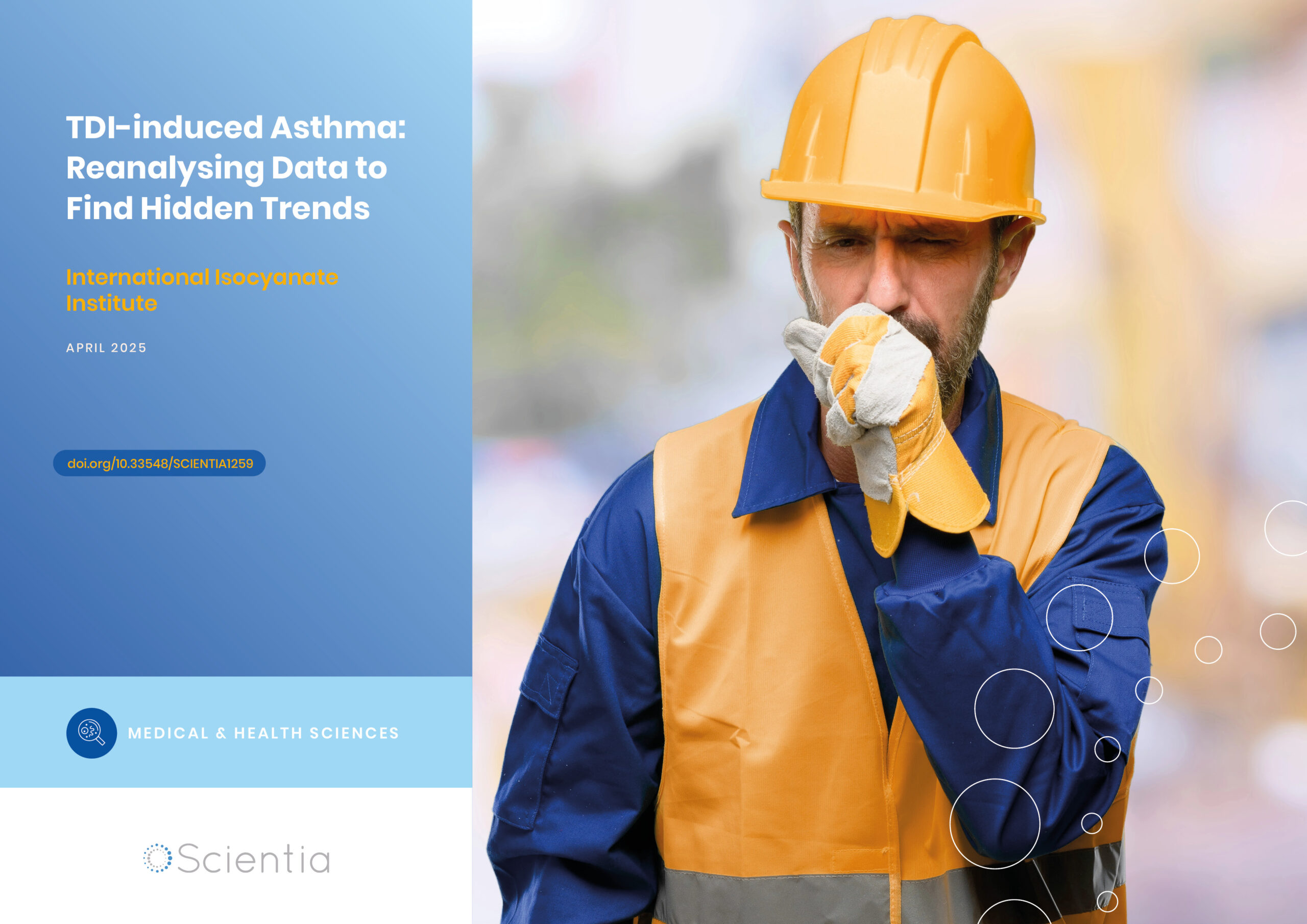
International Isocyanate Institute | TDI-induced Asthma: Reanalysing Data to Find Hidden Trends
Even if you’ve never heard of them, you’ve used polyurethanes. Producing them requires toluene diisocyanates, which may/can induce asthma when inhaled. A 5-year study claimed to conclude that cumulative TDI exposure over time was indicative of asthma incidence. However, a reanalysis by a team at the International Isocyanate Institute points the finger instead at the frequency of unprotected high-exposure events, like accidental spills or plant maintenance. This finding guides the way for future advances in worker safety.

Training Deep Learning AI to Predict microRNA-Gene Interactions
Non-coding microRNAs (miRNAs) have important regulatory functions but are also implicated in various diseases. Mr Seung-won Yoon, PhD candidate at Chungnam National University, Republic of Korea, is training deep learning AI models to predict miRNA-gene associations. His research has implications for understanding disease pathogenesis, particularly cancer, and repurposing drugs for untreatable diseases.
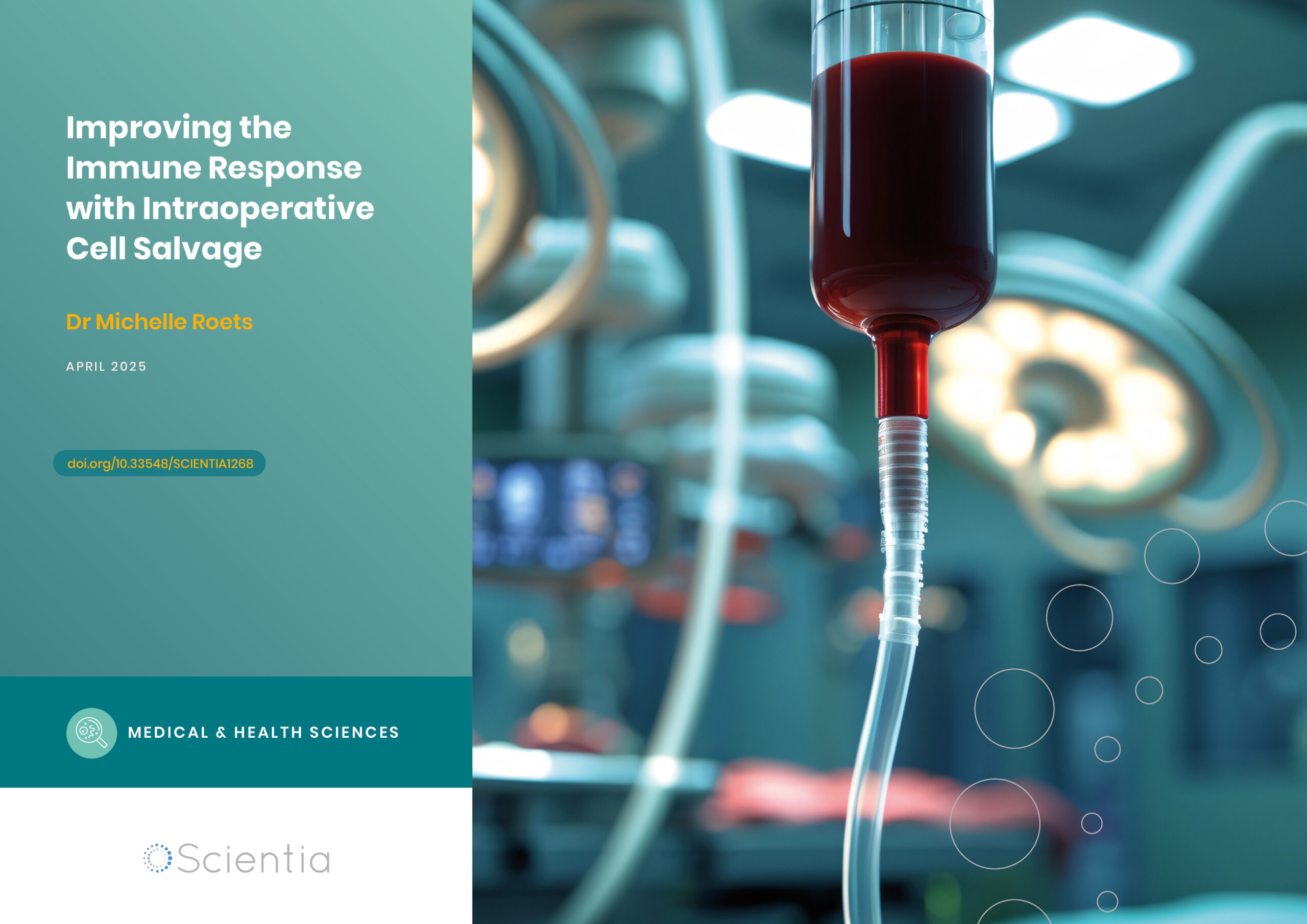
Improving the Immune Response with Intraoperative Cell Salvage
Undergoing surgery comes with many risks. Numerous factors can influence the outcome, from the choice of anaesthesia to the type of operation. Long and complex procedures can require blood transfusions, which introduce yet another risk factor to the mix. Dr Michelle Roets from the Royal Brisbane and Women’s Hospital in Queensland seeks to help mitigate these risks through intraoperative cell salvage, a different type of blood transfusion which could revolutionise surgical outcomes.