
Dr. Eric Buckland – Translational Imaging Innovations: Accelerating Ophthalmic Research Through an Integrated Online Platform
Led by Dr. Eric Buckland, Translational Imaging Innovations, Inc. (TII) provides purpose-driven software systems that drive such ophthalmic research forward. The TII image management platform provides researchers with the tools to manage multifaceted imaging workflows and efficiently organize and analyse complex sets of images and data to accelerate the development of new diagnoses and treatments for eye diseases. By unleashing the power of the eye, TII aims to transform medicine.
The Window to Our Soul and Our Health
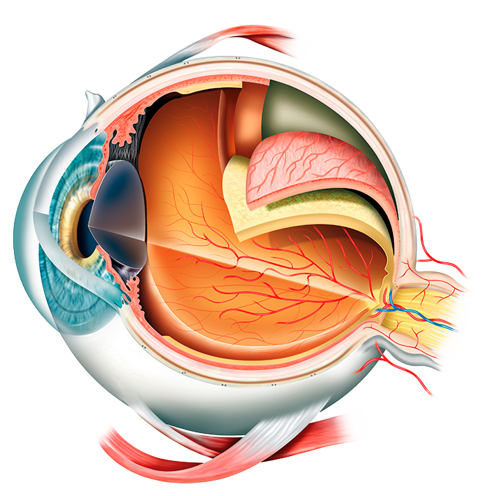
Like a digital camera, the eye captures images of our world by focusing light through the lens at the front of our eye to the sensor – our retina – at the back of the eye. Our retina is part of our central nervous system, circulates blood within our cardiovascular system, and enjoys a privileged role in our immune system. We count on our eyes to interact with the world around us, and in return, our eyes constantly tell us about our health.
Clinicians and scientists rely on advanced imaging technologies for patient care as well as for the development of new treatments to slow down progressive vision loss and prevent blindness. Increasingly, these same ocular imaging tools are making their way into the study of neurological and systemic disease.
Degenerative Eye Disease: Diagnosis and Prognosis
Degenerative eye diseases worsen progressively over time. One of the most common forms is macular degeneration in which an area of the retina (the macula) wears down, leading to vision loss. The exact cause is not well understood, but we believe the same factors that lead to heart disease contribute to it. And much like heart disease, macular degeneration advances with age. Currently, age-related macular degeneration (AMD) is the leading cause of blindness in the Western world and the incidence is expected to rise considerably in the coming years.
Optical coherence tomography (OCT) is a three-dimensional imaging technique that has dramatically improved the management of macular degeneration. OCT provides clear images for the assessment of dry macular degeneration, characterized by deposits of a fatty substance called drusen that builds up in the retina gradually over time. There are currently no treatments for dry AMD but the dry condition may develop into the wet form. Wet macular degeneration is caused by abnormal blood vessels developing in the retina. Although this condition worsens more rapidly, it is treatable. For most people, eye injections of monoclonal antibodies called anti-VEGF prevent the condition from worsening. OCT has played a critical role in the management of wet AMD, providing immediate insight that the clinician uses to determine the timing of eye injections to preserve sight, maximize patient comfort, and control costs.
Glaucoma is a degenerative eye disease that is caused by obstructed flow and drainage of fluids that circulate through the eye. This obstructed flow increases the pressure inside the eye, causing damage to the optic nerve which connects the eye to the brain. Loss of this nerve function slowly results in vision loss in the peripheral visual field and blindness if left untreated. Unfortunately, the early stages of glaucoma do not present with symptoms. While current estimates suggest more than 3 million Americans have glaucoma, only half are thought to have received a diagnosis. Without a diagnosis and in the absence of treatment, the condition continues to irreversibly progress.
Glaucoma is typically identified before symptoms develop at routine optician visits and is confirmed through various tests. These can include an eye pressure test, a gonioscopy, a visual field test, or an optic nerve assessment that uses optical coherence tomography. Glaucoma remains a significant medical challenge, and advanced imaging technologies are in constant development to improve decision-making in glaucoma management.
Inherited retinal diseases (IRDs) are caused by the mutation of at least 1 of more than 270 associated genes. As a group, IRDs are diverse with some leading to a gradual loss of vision over time and others resulting in vision loss much earlier, such as in infancy or young adulthood. Although relatively rare, IRDs are the leading cause of blindness in people aged 15 to 45, affecting 1 in every 2,000 individuals. The effective diagnosis and tracking of the progression of IRDs in individuals requires the identification of sensitive and specific biological markers, or in simpler terms, measures of a biological or pathogenic state.
This can be achieved using an advanced imaging technique called adaptive optics (AO), enhanced retinal imaging that allows for a direct view of photoreceptors in the retina. AO retinal imaging is increasingly being used to support gene research into genetic vision loss and as a tool to support the development of gene therapy. AO imaging shows how photoreceptors (rods and cones) are distributed in the eye. While two patients may present the same clinical conditions, the pattern of rods and cones may differ significantly. The analysis of these differences may prove essential in determining the type of genetic defect and in assessing the performance of new gene therapies.
Even More Than Meets the Eye
Eye examinations can reveal much more than just diseases of the eye. Systemic diseases like diabetes and even Alzheimer’s disease can also be detected through the examination of eye damage. For example, diabetic retinopathy occurs as a result of high blood sugar levels seen in diabetes. The retina requires a constant supply of blood, but if the blood is full of sugar, the vessels that transport it become damaged and the retina becomes impaired, potentially leading to retinal detachment, vision loss, and blindness without intervention. With regard to Alzheimer’s, direct diagnosis is not yet fully functional, but research suggests that observing a diminished density of blood vessels in the retina can be a signal of neurodegenerative disease.
Researchers and innovative young companies are using the eye-brain connection to study neuromuscular diseases such as multiple sclerosis. Small involuntary motions of the eye, called micro-saccades, are impacted by neurological disease progression. These micro-saccades can now be extracted through precision tracking of retinal motion using imaging technologies. Artificial Intelligence (AI) methods to analyse the motion used to monitor slight changes in saccades associated with neuromuscular disease progression are opening a new field in diagnosis of central nervous system disease through non-invasive imaging of the eye.
Translational Imaging Innovations, Inc.
As demonstrated above, eye imaging is essential in the identification and management of pathologies directly and indirectly related to visual impairment. AI is growing in importance for the analysis of images. However, systems developed to use images for clinical patient care lack the coherence and organization required for more advanced research.
Dr. Eric Buckland is overcoming these limitations and transforming the field of ophthalmic research through his company, TII. Dr. Buckland explains, ‘Our mission is the provision of better diagnostics and better therapies that provide more predictable benefits to patients faster, at a lower cost, and with less frustration.’
Before TII, Dr. Buckland was the CEO and Co-founder of Bioptigen. This company enhanced the use of optical coherence tomography by dramatically improving image quality and increasing system flexibility. Bioptigen innovations have made a lasting impact in early-stage ocular research and pediatric ophthalmology. Bioptigen was the first to introduce real-time OCT imaging of the retina and the cornea into ocular surgery. Bringing a new class of imaging to researchers and clinicians, Bioptigen manufactured and distributed their advanced OCT machines around the world, improving the use of imagery for both research and patient care.
Through this early work, Dr. Buckland identified the ‘tremendous untapped potential to extract latent medical information from the exquisite images we can acquire through the eye.’ Building on this, Dr. Buckland is now creating innovative and marketable software solutions to fulfill unmet ophthalmic needs through TII.

An Online Platform for Accelerating Ophthalmic Research
Quantitative biomarkers are needed to fully utilize the retinal images from techniques like optical coherence tomography for clinical diagnosis. To facilitate research into finding these biomarkers, the team at TII created an online, integrated platform to collect, store, share, and manage ocular images and data. This center, called Lattice, allows researchers to access and analyse considerably more information than before.
AI is becoming fundamental to medical advancements. Complex algorithms process huge amounts of images and data to automatically identify patterns related to targeted disease or condition. In ophthalmology, AI can be used to identify disorders of the eye by essentially comparing a new patient’s scan to its database of known pathologies. AI-enabled techniques for automated diagnoses of diabetic retinopathy using ocular imaging, for example, were the first medical AI solutions to be cleared by the FDA. The field of AI in medical imaging is still in its infancy. AI requires the systematic management and analysis of large numbers of images validated by experts, to uncover ‘explainable’ patterns in images to guide effective clinical interpretation.
Dr. Buckland’s work means that annotated images from all over the world can be stored in a distributed but uniform manner, providing remote accessibility to large volumes of images and data that for AI programs to work with. TII systems are designed to make data access easier, more transparent, and less frustrating. TII enables the free flow of actionable ophthalmic information and empowers researchers to develop devices and therapeutics faster and at a lower cost. The goal is faster, more reproducible results and improved communication and collaboration across the field of ophthalmic research. Ultimately, this may save the eyesight and improve the overall health of millions of people.
Ensuring the Software is User-friendly
The Translational Imaging Innovations platform is supported by four pillars. Lattice is an easy-to-use Web application that incorporates electronic record keeping, protocol adherence, scheduling, subject and exam tracking, and team communications. Lattice is an information system built around our proprietary data model: our Data Genome. ocuvault™ is a unique data transport and storage system for the secure transfer of images and data. ocuVault maintains data provenance and security, ensuring accessible, actionable, and auditable data throughout a project lifecycle. oculink™ is a visual software solution that aggregates data and images for rapid curation, visualization, and annotation, designed by researchers for researchers. Mosaic is a work environment for studying the impact of disease and therapeutics on photoreceptors. Mosaic was originally developed by Dr. Robert Cooper and Dr. Joseph Carroll at the Dennis P. Han, MD Advanced Ocular Imaging Program (AOIP) at the Medical College of Wisconsin (MCW).
In addition, TII is licensed to the AOIP Image Bank, which houses AO and OCT images and data from more than 1,800 real patients with one or more degenerative retinal. TII continues collaboration with the Advanced Ocular Imaging Program (AOIP) at MCW and the Ocular and Computer Vision Laboratory at Marquette University to research imaging biomarkers of inherited retinal disease.
Real-world Applications
The National Eye Institute (part of the National Institutes of Health in Washington, DC) has recognized the capabilities and vision of TII and has awarded grants to aid in the continued development of its initiatives. TII software is already used for managing 80 clinical research projects. Mosaic is integrated into an international research project sponsored by the Foundation Fighting Blindness®. ocuVault and ocuLink are in early deployments through relationships with innovative imaging system startups, ophthalmic biophysics research collaborators, and drug development organizations. As these programs continue to be utilized, their impact will become more recognized and widespread.
Through his dedicated work, Dr. Buckland is truly transforming ophthalmic research. Lattice, Mosaic, and ocuVault and ocuLink promise to become invaluable tools for finding and improving new quantitative biomarkers for degenerative eye diseases. By creating a dedicated space that pulls together images and data and combines them with AI to push forward diagnostics, Dr. Buckland is facilitating the future of eye imaging research. Now—and in the coming years—these platforms will be used to improve diagnoses and therapies so that people suffering from degenerative eye disease will benefit from dramatically improved health outcomes.
SHARE
DOWNLOAD E-BOOK
LISTEN TO THE AUDIO
REFERENCE
https://doi.org/10.33548/SCIENTIA769
MEET THE RESEARCHER

Translational Imaging Innovations, Inc.
Hickory, NC
USA
Dr Eric Buckland completed his Bachelor and Master of Science degrees in Physics at North Carolina State University in Raleigh. He went on to receive his PhD in Optics from the University of Rochester’s Institute of Optics in New York state. Throughout his impressive career, Dr Buckland has taken on a variety of leadership positions in global science innovation companies. As an entrepreneur, Dr. Buckland co-founded Bioptigen, which he managed from inception to exit. Currently, Dr Buckland is the Chief Executive Officer and Founder of Translational Imaging Innovations, situated in Hickory, North Carolina. He has assembled a great team to work with great collaborators on developing the next generation of software systems for discovering, developing, and deploying new markers of disease through the window of the eye. His passion is enabling his community to deliver the benefits of their research to patients, faster, at lower cost, and with better outcomes. Dr. Buckland is a Fellow of the American Institute of Medical and Biological Engineering (AIMBE) and a Senior Member of Optica.
CONTACT
E: eric@tiinnovations.com
W: https://www.linkedin.com/in/buckland/
FURTHER READING
https://sbir.nih.gov/statistics/success-stories/bioptigen
![]()
REPUBLISH OUR ARTICLES
We encourage all formats of sharing and republishing of our articles. Whether you want to host on your website, publication or blog, we welcome this. Find out more
Creative Commons Licence (CC BY 4.0)
This work is licensed under a Creative Commons Attribution 4.0 International License. 
What does this mean?
Share: You can copy and redistribute the material in any medium or format
Adapt: You can change, and build upon the material for any purpose, even commercially.
Credit: You must give appropriate credit, provide a link to the license, and indicate if changes were made.
SUBSCRIBE NOW
Follow Us
MORE ARTICLES YOU MAY LIKE
Dr Yong Teng | Improving the Outlook for Head and Neck Cancer Patients
Dr Yong Teng at the Emory University School of Medicine is working with colleagues to overcome the high mortality of individuals diagnosed with cancers affecting the head and neck. One of his approaches is based on understanding the particular mechanisms of the ATAD3A gene, which new insights suggest are closely related to cancers affecting the head and neck.
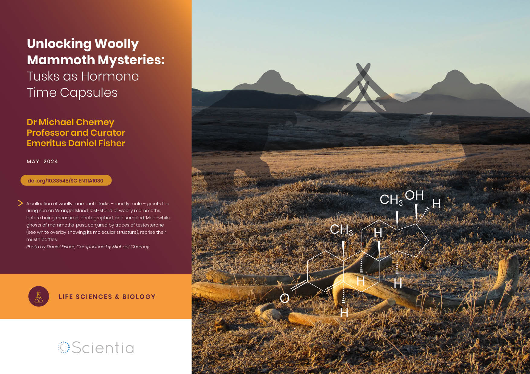
Dr Michael Cherney – Professor Daniel Fisher | Unlocking Woolly Mammoth Mysteries: Tusks as Hormone Time Capsules
The impressive tusks found on proboscideans (the order of mammals that includes elephants, woolly mammoths, and mastodons) are like time capsules, preserving detailed records of their bearers’ lives in the form of growth layers and chemical traces. Frozen in time for thousands of years, these layers can unlock secrets about the lives of long-extinct relatives of modern elephants. Dr Michael Cherney and Professor Daniel Fisher from the University of Michigan used innovative techniques to extract and analyse steroid hormones preserved in woolly mammoth tusks. This ground-breaking work opens new avenues for exploring the biology and behaviour of extinct species.
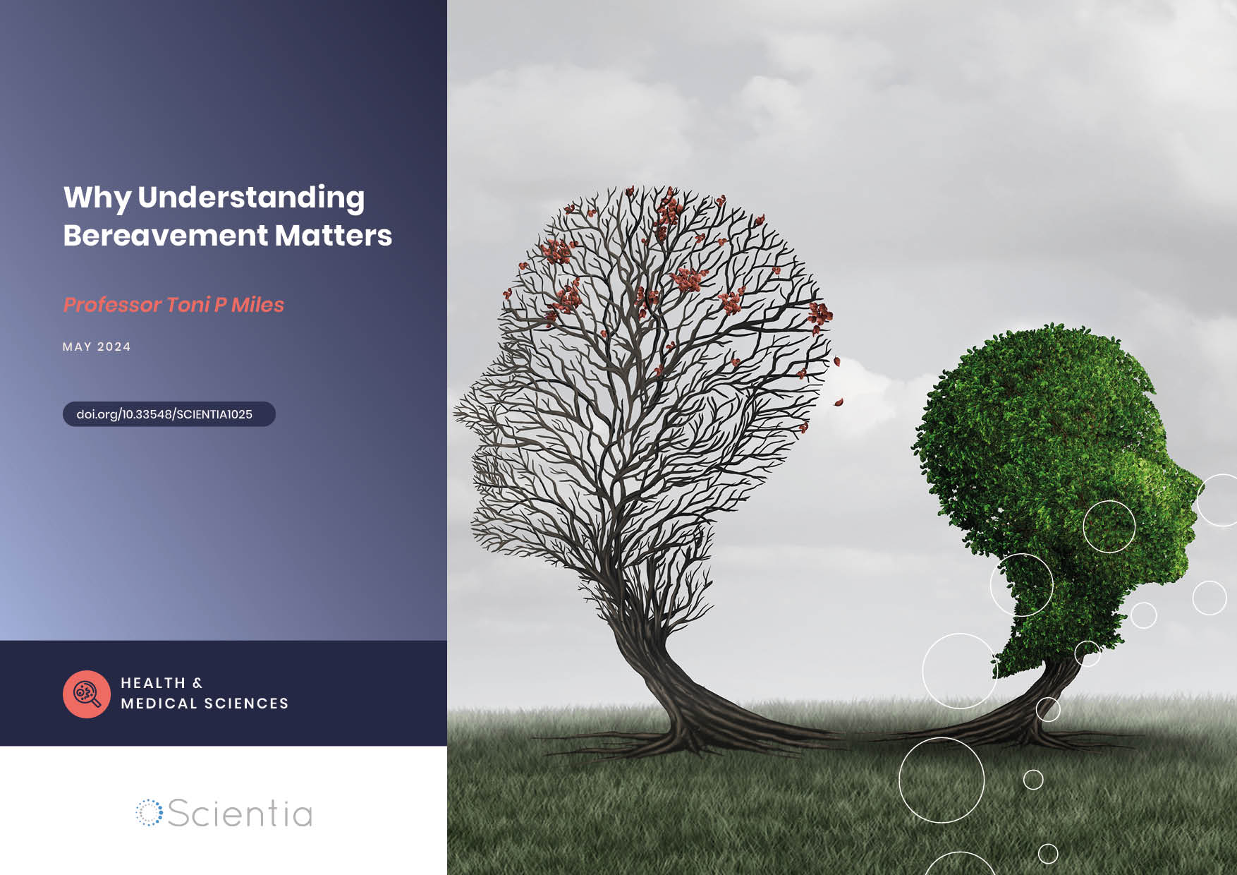
Professor Toni Miles | Why Understanding Bereavement Matters
Professor Toni Miles has dedicated her research efforts to measuring bereavement and its impact on population health. Individual experience with bereavement is commonplace, but we know little about its impact on society when there is an instantaneous experience by a large number of individuals, i.e., mass bereavement. To measure its occurrence, her research with colleagues first confirmed that bereavement can be effectively measured in population surveys. Professor Miles argues that we should use such approaches to deliver interventions aiming to reduce the negative consequences of bereavement on individuals. By measuring bereavement in communities, these data become a cost-effective way to increase resilience, reduce demands on healthcare systems, and enhance public safety.
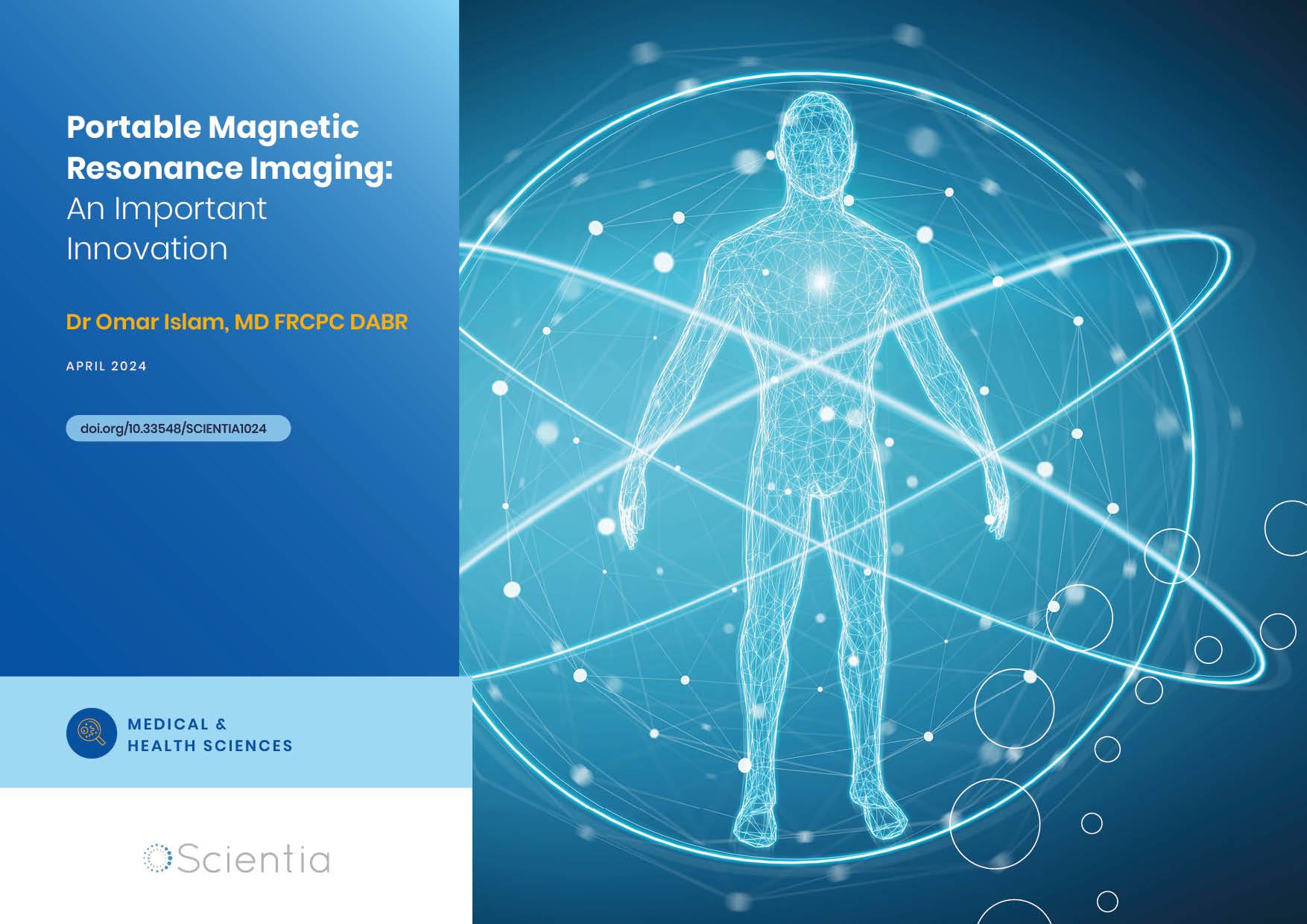
Dr Omar Islam | Portable Magnetic Resonance Imaging: An Important Innovation
Imaging technologies are vital in modern medicine and have revolutionised how clinicians make diagnoses and monitor disease progression. However, the necessary equipment – such as a scanner for magnetic resonance imaging (MRI) – is very large and expensive, requiring patients to go to the scanner rather than receiving scans as bedside care. This takes up valuable staff time and resources, and can present further risks to patients. Dr Omar Islam from Queen’s University and Drs Aditya Bharatha and Amy Lin from the University of Toronto are showing how portable MRI scanners may offer a viable alternative that benefits patients and healthcare systems.