
Professor Andrew Adey | High-Throughput Analysis of DNA Methylation at the Single-Cell Level
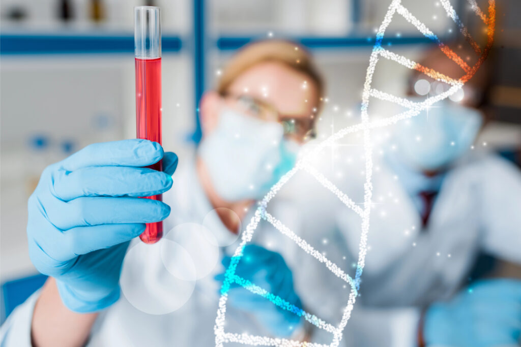
DNA methylation is a key epigenetic process. Conventional methods for analysing methylation have been cumbersome or technically unfeasible. Professor Andrew Adey at Oregon Health and Sciences University is developing high-throughput workflows that analyse the methylome with single-cell granularity.
Epigenetic Implications of DNA Methylation
Our phenotypes are the result of both our genes and environment. This interplay is turning out far more complex than classical genetics would suggest. We now know that hereditary phenotypes can occur without changes to DNA sequence. The field of epigenetics is concerned with investigating how and why this happens.
A key epigenetic process is DNA methylation – the addition of methyl groups to DNA, usually catalysed by DNA methyltransferase enzymes. This commonly occurs at the 5-position of cytosine (C) nucleotides, where a C is followed by G in the 5’–3’ direction, known as CpG sites. CpG islands are genomic regions rich in CpGs. In humans, CpG islands often overlap with gene promoters, where methylation leads to gene silencing by repressing transcription. The nature of DNA methylation, as a chemical modification to the DNA itself, makes it the most basal layer of gene regulatory control. In a sense, the patterning of DNA methylation acts as the first filter on the genome to enable cells to fine-tune their programs and carry out their specified function.
Environmental conditions may influence changes in methylation and are a feature of physiological development, cell differentiation, and ageing. Conversely, aberrant methylation status has been implicated in various diseases. In cancer, the genome frequently loses methylation, resulting in a hypomethylation phenotype that allows cancer cells to explore a broader space of gene programs, fuelling the cancer’s ability to evolve and evade treatment. Curiously, methylation patterns in gametes may be passed to offspring, raising the possibility of epigenetic inheritance.
Conventional Approaches to Analysing Methylation
The ‘methylome’ can vary depending on tissue type, developmental stage, ageing, disease status, or cancer. Within each tissue, the methylation patterns can vary extensively between cells. This diversity is at its peak in the brain, where neurons exhibit thousands of specialised patterns. Thus, methylation assays with single-cell granularity are needed to enable cell type-specific characterisation. Single-cell assays that assess mRNA or chromatin accessibility have already been established. However, methylomics has lagged behind advances in transcriptomics or chromatinomics, largely due to technical limitations.
The primary method of deciphering the patterns of DNA methylation in the genome is to leverage the chemical Sodium bisulphite or enzymatic treatments. Each of these unmethylated C bases is converted to uracil and then sequenced as thymine. Meanwhile, methylated Cs retain their identity. After sequencing, observing where the Cs and Us are can indicate the presence or absence of methylation.
The challenge this poses to single-cell assays is that it requires a multi-step process involving DNA denaturation, multiple wash steps, and buffer exchanges – highly cumbersome and not conducive to high-throughput analysis. Alternative single-cell methods based on a microwell plate format are available. This is a ‘one-pot’ process – individual cells or nuclei are isolated and undergo bisulphite or enzymatic reactions in a single well. This is scalable if multiple plates and robotics are involved. Nevertheless, it is still expensive and technically challenging, rendering it impossible for most research laboratories.
A New Approach to Single-Cell Methylation Profiling
Professor Andrew Adey and his group at Oregon Health and Sciences University have devised advanced methylation profiling methods that circumvent the technical limitations of established assays.
They previously developed a high-throughput workflow, named sciMETv2, an upgraded iteration of a single-cell workflow known as sciMET. In sciMETv2, cell nuclei are fixed, and nucleosomes (DNA-histone chromatin complexes) are disrupted, releasing the DNA. The nuclei are then ‘barcoded’ via tagmentation in a 96-well plate, with about 5,000 nuclei per well.
Tagmentation, a technology pioneered by Professor Adey as a graduate student, involves transposomes, which are complexes of a transposase enzyme loaded with adapter oligos. Adapters contain a double-stranded sequence recognised by the transposase and a single-stranded 5’ overhang containing a DNA sequence ‘barcode’. In tagmentation, the adapter overhangs covalently bind the nuclear DNA. The post-tagmentation nuclei are pooled together and sorted via flow cytometry into 96-well plates, with 15 to 30 nuclei per well. This low number is important – it is unlikely that two nuclei with the same tagmentation index ‘barcode’ end up in the same well. The nuclei then undergo bisulphite conversion within each well, before being cleaned up. The library molecules are then modified to add a final sequencing adapter and indexed again during amplification. This process allows for thousands of single-cell methylation libraries to be produced in a relatively simple and cost-effective experimental setup.
Professor Adey’s group devised two variations of DNA library preparation following bisulphite conversion – sciMETv2.LA (linear amplification) and sciMETv2.SL (splint ligation). With sciMETv2.LA, DNA libraries are subjected to multiple rounds of adapter extension, giving multiple opportunities to capture each library fragment at the cost of a longer workflow. Alternatively, in sciMETv2.SL, the library is processed using a direct adapter ligation, giving only one shot to capture each fragment but wrapped in a rapid and cost-effective protocol. After either method, the libraries undergo qPCR, followed by NGS. Overall, sciMETv2.LA produces higher coverage of the methylome per cell, achieving as high as 50% of CpG sites versus sciMETv2.SL, which provides less coverage yet is sufficient for the large majority of applications.
Reducing Sequencing Burden with Target Enrichment
Although sciMETv2 is conducive to high-throughput analysis, one limiting factor remains – sequencing. Both sciMETv2.LA and sciMETv2.SL provide genome-wide access, with a typical read count between 2 to 6 million reads per cell. This can be expensive and time-consuming.
Professor Adey’s group have developed a method of target enrichment known as sciMET-cap to capture sufficient information while reducing sequence reads. This deploys an off-the-shelf 125 Mbp panel (from Twist Biosciences) that selectively captures regulatory sequences. sciMET-cap can be applied to DNA libraries generated by sciMETv2.LA or sciMETv2.SL. Libraries are pooled together and run through the panel. DNA with methylated regions complementary to the panel are hybridised and captured. Thermocycling at 95°C and 60°C is needed for efficient hybridisation, and non-specific hybridisation is avoided by adding blocker oligos. Subsequently, the hybridised library mixture is bound to streptavidin beads, amplified with PCR, and undergoes NGS.
Professor Adey’s group have carried out experiments to optimise the performance of sciMET-cap. They investigated the methylomes of human peripheral blood mononuclear cells and the middle frontal gyrus region of the human brain. Using a peripheral blood mononuclear cell library, they explored various capture conditions – standard or custom blockers, and 63°C or 67°C wash temperature for binding streptavidin beads. They found that the capture of differentially methylated loci was consistent across conditions, indicating that standard blocking and wash conditions are sufficient.
Producing Large Methylome Datasets
The group put the high-throughput potential of sciMET-cap to the test with large methylome datasets sequenced using a standard benchtop sequencer. They produced a sciMETv2.SL-derived library dataset from 5,952 PBMC cells. By comparing methylation status at promoters of marker genes against known profiles, they could assess different cell types – monocytes, B-cells, natural killer cells, and T-cells, with clusters for CD4-expressing and CD8-expressing cells. sciMET-cap target enrichment led to a 33-fold improvement in identifying differentially methylated regions (195,483 vs 5,914) from 1.7 billion war sequence reads. Similarly, they produced a sciMETv2.LA-derived library of 6,123 gyrus cells from the human cerebral cortex.
By assessing methylation patterns at marker gene promoters, they identified 12 clusters consisting of astrocytes, microglia, oligodendrocytes, excitatory neurons, and inhibitory neurons. Per-cluster genomic coverage was relatively low, suggesting that increased sequencing depth may be needed for highly heterogeneous tissues. Nevertheless, sciMET-cap enabled them to sequence 1.2 billion raw reads, equating to 200–275 thousand raw sequence reads per cell, identifying 64,774 differentially methylated regions – a truly impressive feat!
These experiments demonstrate what is possible on a modest budget and within the range of what a typical research programme would aim for in a single-cell experiment. While the cost of sciMET-cap experiments still exceeds that of single-cell transcriptomics or chromatin assays, it brings it within the same ballpark.
Implications for Epigenetics Research
As the field of epigenetics comes of age, there is a need for the development of robust methylomics technologies. Professor Adey’s group are at the forefront of this, with the goal of ‘democratising single-cell methylomics’. Their assay, sciMETv2, can generate single-cell methylomes at 10-fold higher throughput and 10-fold lower cost. Their target enrichment method, sciMET-cap, reduces sequencing costs by 10-fold. These innovations could make single-cell DNA methylation profiling a reality for labs, where previously this was impossible. This opens the door to elucidating the epigenetic implications of methylation in the context of health and disease, physiological development, ageing, and gene regulation.
SHARE
DOWNLOAD E-BOOK
REFERENCE
https://doi.org/10.33548/SCIENTIA1076
MEET THE RESEARCHER

Professor Andrew C. Adey
Molecular & Medical Genetics, School of Medicine
Oregon Health & Sciences University
Portland, OR
USA
Andrew Adey is Professor of Molecular and Medical Genetics at Oregon Health & Sciences University (OHSU). His biotechnology journey began at the University of Texas, Austin. Here, he obtained a BS in Biochemistry in 2008 and served as Interim Director of the Microarray Core Facility. Subsequently, he embarked on his doctoral studies at the University of Washington, gaining his PhD in Molecular and Cellular Biology in 2014. He joined OHSU as an assistant professor in 2014, then progressed to associate professor and finally, tenured professor. His broad research interests include epigenetics, genomics, single-cell ‘omics’ technologies, and computational biology, focusing on cancer and neurological disorders. He has a special interest in developing high-throughput single-cell methods for genomic and epigenetic analysis. Keen to impart his insights to the next generation of scientists, he teaches courses at OHSU on epigenetics, single-cell omics, and advanced genome sciences.
CONTACT
W: https://www.ohsu.edu/people/andrew-c-adey-phd , www.adeylab.org
X: @acadey80
KEY COLLABORATORS
Brian J O’Roak, Molecular and Medical Genetics, Oregon Health and Science University
Kevin M Wright, The Vollum Institute, Oregon Health & Science University
Rosalie C Sears, Molecular and Medical Genetics, Oregon Health & Science University
Arpiar Saunders, The Vollum Institute, Oregon Health & Science University
Gail Mandel, The Vollum Institute, Oregon Health & Science University
Ted Braun, Division of Hematology/Medical Oncology, Oregon Health & Science University
Gurkan Yardimci, Knight Cancer Institute, Oregon Health and Science University
FUNDING
National Institutes of Health
National Cancer Institute
National Institute of Mental Health – The Brain Initiative
National Institute of Child Health and Human Development
Silver Family Innovation Fund
FURTHER READING
SN Acharya, RV Nichols, LE Rylaarsdam, et al., sciMET-cap: High-throughput single-cell methylation analysis with a reduced sequencing burden, bioRxiv, 2023, 2023.07.12.548718. DOI: https://doi.org/10.1101/2023.07.12.548718
RV Nichols, BL O’Connell, RM Mulqueen, et al., High-throughput robust single-cell DNA methylation profiling with sciMETv2, Nature Communications, 2022, 13(1), 7627. DOI: https://doi.org/10.1038/s41467-022-35374-3
REPUBLISH OUR ARTICLES
We encourage all formats of sharing and republishing of our articles. Whether you want to host on your website, publication or blog, we welcome this. Find out more
Creative Commons Licence (CC BY 4.0)
This work is licensed under a Creative Commons Attribution 4.0 International License. 
What does this mean?
Share: You can copy and redistribute the material in any medium or format
Adapt: You can change, and build upon the material for any purpose, even commercially.
Credit: You must give appropriate credit, provide a link to the license, and indicate if changes were made.
SUBSCRIBE NOW
Follow Us
MORE ARTICLES YOU MAY LIKE
Dawn Dunbar | Harnessing Machine Learning to Enhance Diagnosis of Feline Infectious Peritonitis
Feline infectious peritonitis (FIP) is a severe and often fatal viral disease of cats which poses significant diagnostic challenges for veterinarians. Dawn Dunbar from the University of Glasgow is leading a research study with the goal of applying machine learning to revolutionise the diagnosis of FIP. By leveraging routinely collected clinical laboratory data, this innovative approach may pave the way for more accurate and timely diagnoses, ultimately improving outcomes for affected cats and their owners.

Professor Irene Díaz-Moreno – Professor Miguel A. De la Rosa | The Diverse Interactome of Cytochrome c: Beyond Respiration
All living things are comprised of cells, and to function, most of them use oxygen to break down food molecules to obtain chemical energy, a process known as cell respiration. Critical to this is the macromolecule cytochrome c, but this redox haemoprotein also boasts a diverse set of functions beyond respiration. Professor Irene Díaz-Moreno and Professor Miguel A. De la Rosa, both leading members of cicCartuja’s Biointeractomics Research Group at the University of Seville, are using cutting-edge investigational tools to study the full ‘interactome’ of this multifunctional molecule.
Dr Wolfgang Quapp – Professor Josep Maria Bofill | Shaping Reactions: The Exciting World of Mechanochemistry and Molecular Interactions
Dr Wolfgang Quapp and Professor Dr Josep Maria Bofill from the University of Leipzig and Universitat de Barcelona, respectively, are leading voices in the newly emerged sector of mechanochemistry. Their fascinating work reveals how external forces can manipulate molecular behaviour and influence chemical reactions.
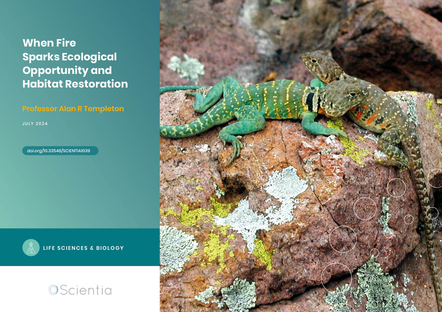
Professor Alan Templeton | When Fire Sparks Ecological Opportunity and Habitat Restoration
How far would you be willing to go to save an endangered species? Would you consider burning part of a forest as a solution? As unconventional as it may sound, conservationists sometimes resort to such measures to restore lost habitats. One remarkable example is the efforts to save eastern collared lizards – and indeed the entire biological community in which they live – in the Ozarks, spearheaded by American geneticist and statistician Professor Alan Templeton of Washington University in St Louis, USA.