
Dr Frank Wuest – Innovative Strategies for Imaging Cancers
Dr Frank Wuest is the Director of the Division of Oncologic Imaging at the University of Alberta, Canada, where he holds the Dianne and Irving Kipnes Chair in Radiopharmaceutical Sciences. His main research interests revolve around the multidisciplinary field of cancer imaging but he is particularly interested in the possibilities of translating techniques developed in the laboratory to clinical settings to enhance patient care. In particular, he focuses on the design, synthesis, and validation of novel molecular probes to optimise the current diagnosis and treatment of cancer.
The aim of Dr Frank Wuest’s research at the University of Alberta, Canada, is to dig deeper into the use of molecular imaging techniques to detect cancer. His ultimate goal is to facilitate the translation of novel biomarkers, which have diagnostic and therapeutic potential, from the laboratory to clinic. Biomarkers are defined as characteristics that can be measured from imaging data as indicators of biological processes, including diseases. In addition to his work on cancer imaging, Dr Wuest has also been involved in investigating how cancer cells change to adapt to different conditions, such as low oxygen.
Particular attention is given to the use of novel positron emission tomography (PET) radiopharmaceuticals and pre-clinical PET imaging technology. PET imaging is a technique that can be used to observe metabolic and molecular processes in the body and is often used for diagnosing diseases.
The process works by detecting pairs of gamma rays, which originate from the collision of a positron, emitted by positron emitters, with an electron. The most common positron-emitting radionuclide is fluorine-18 (18F). Radionuclides like 18F are introduced into the body attached to biologically active molecules called radiotracers, and it is the accumulation of these radiotracers in certain tissues and organs which can be turned into a 3D image to reflect their spatial and temporal biodistribution in vivo.
The radiotracer is normally associated with a specific biological process, such as glucose uptake by cells, and can, therefore, be used to infer information about tissue metabolic activity. As Dr Wuest explains, ‘The availability of innovative PET radiotracers to visualise and study biochemical processes in living organisms is an important driving force and a key element for the successful application of PET imaging technology in biomedical and clinical research.’
In the 15 years since becoming an independent researcher, Dr Wuest and his group have pioneered the development of novel chemistry techniques for molecular imaging and the validation of novel PET radiotracers and PET imaging assays that are associated with human pathologies. These assays include the molecular imaging of cancer, metabolic diseases, and neuroreceptors (which are receptor molecules activated by neurotransmitters).

Click Chemistry and COX-2
Part of Dr Wuest’s current research is dedicated to the design and validation of probes for molecular imaging of COX-2, which is an important biomarker in cancer and inflammation. His efforts were rewarded by the development of the first successful PET imaging probe ([18F]pyricoxib) for targeting COX-2 in cancer.
Leading on from this, Dr Wuest’s group has also pioneered the development of fluorescent-labelled probes for the molecular imaging of COX-2 and were the first to use click chemistry for the design of highly specific and potent COX-2 inhibitors. Click chemistry is a class of biocompatible small molecule reactions to generate new compounds. This is often done between a biomolecule and a molecule containing a radionuclide (as for PET imaging). These reactions normally produce rapidly the new molecule in high yield with high specificity. This makes click chemistry ideal for targeting molecules in complicated biological environments, including in living organisms. Dr Wuest’s team was among the first to apply click chemistry to the radiolabelling of peptides with 18F.
Even more excitingly, the team used in situ click chemistry for the preparation of highly potent and selective COX-2 inhibitors, where the COX-2 binding site was used as a molecular template to generate its own inhibitors. This removed the need for laborious synthesis and screening of a range of compounds, which is the usual approach for drug discovery. The methods used in the study described above have the potential to be extrapolated for economical and fast screening of other possible drug targets. Dr Wuest’s team is currently investigating how the technique could be expanded to detect COX-2 expression at the cellular and whole-body level.
Various 18F-labelled peptides have been designed by Dr Wuest and his collaborators and these molecules can be used as radiotracers to target a wide range of biomarkers associated with cancer and inflammation. The group has also worked with a different short-lived positron emitter, 11C. Together with 18F, the expansion of 11C- and 18F-labeled radiotracers has enabled Dr Wuest’s research group to investigate site-specific labelling techniques to incorporate the radionuclide into a specific position of a given molecule. Other, non-standard PET radionuclides have also been investigated in order to determine whether they may be useful for the molecular imaging and therapy of cancer.
‘The availability of innovative PET radiotracers to visualise and study biochemical processes in living organisms is an important driving force and a key element for the successful application of PET imaging technology in biomedical and clinical research.’

Breast Cancer
Breast cancer is the most common cancer affecting women worldwide. As cancer cells proliferate rapidly, there is often limited oxygen available to the cells resulting in changes in molecular processes linked to low oxygen and hexose transport. These hexose transport molecules can be targeted by radiotracers and it has been suggested that low oxygen conditions indicate a poor prognosis for therapy outcomes and survival in patients with breast cancer.
Dr Wuest and his team investigated the expression of these hexose transport molecules in different cancer cells grown in the laboratory under low oxygen conditions. Identification of specific metabolic markers on tumour cells will allow a ‘molecular fingerprint’ to be established which can subsequently guide the choice of imaging probe used for diagnosing and treating the different forms of breast cancer.
Taking a slightly different approach, Dr Wuest and his team have also recently started to develop a molecular imaging assay for autotaxin, an enzyme also thought to be associated with chronic inflammation, including cancer. This may also have a particular impact on the treatment of breast cancer patients undergoing radiation therapy.

Prostate Cancer
Prostate cancer is the fifth leading cause of cancer-related death in men in Western countries. A molecule called prostate-specific membrane antigen (PSMA) is often increased in prostate cancer. Previously, PET imaging of PSMA with 18F-labelled radiotracers had several limitations, including the amount of time and effort required in producing 18F-labelled PSMA inhibitors.
Using an automated synthesis would remove this limitation – and this is what Dr Wuest and his team set out to do. They were able to simplify the synthesis of clinically important radiotracer [18F]DCFPyL, using a single step radiofluorination procedure in an automated synthesis unit. In addition, [18F]DCFPyL is taken up by tumours and effectively cleared from the body, preventing unnecessary accumulation of the radiotracer in most organs and tissues of the human body. Pre-clinical results confirmed that this molecule is a promising option for targeted molecular imaging of PSMA in prostate cancer.
In addition, work by Dr Wuest and others at the Washington University School of Medicine tested the ‘serve and protect strategy’, which suggests that administering enzymes at the same time as radiolabelled peptides can increase the survival of the peptides, may not be valid for peptides which naturally show metabolic stability in animal models. Other research projects undertaken by Dr Wuest include the development and clinical translation of radiolabelled and metabolically stabilised peptides for molecular imaging and therapy of breast and prostate cancer. In particular, he has already developed a molecule called BBN2 which, when linked to 18F, may be important for looking at certain types of tumour in breast and prostate cancers.

Ovarian Cancer
The lack of clear symptoms, combined with the high incidence of recurrence, means that ovarian cancer has only a 44.6% five-year survival rate. Cancer antigen 125 (CA125) is a glycoprotein that is overexpressed on the membrane of ovarian cancer cells and may act as a biomarker for ovarian cancer. However, normal levels of the protein can still be associated with the presence of disease. Current assays may be limited in their ability to detect early-stage cancers.
A process called immuno-PET, a version of PET imaging which uses an antibody linked to a radionuclide has shown promise as a strategy that unifies the specificity of antibody binding to a target on the tumour cell with the sensitivity of detection via positron-emitting radionuclides. Dr Wuest and colleagues at MSKCC produced a radiotracer based on an antibody for CA125, which could be used for molecular imaging of the glycoprotein in the body. Furthermore, using a radionuclide to label the antibody for immuno-PET imaging did not compromise the ability of the antibody to bind to its target.
Future Directions
The novel imaging assays that Dr Wuest is involved in designing are vital contributions to cancer care. Specifically, the ‘precision oncology’ concept aims to enhance patient care and outcome through early, more accurate diagnosis and better treatment regimens specifically tailored to each individual patient’s requirements.
Ultimately, the more improvements that can be made to cancer imaging, the better the outcome for the patient. It is this seemingly simple statement that makes the research of Dr Wuest and his colleagues so important for the future of cancer diagnosis and treatment.
Reference
https://doi.org/10.33548/SCIENTIA438
Meet the researchers

Dr Frank Wuest
Department of Oncology
University of Alberta
Edmonton, Alberta
Canada
Dr Frank Wuest is the Director of the Division of Oncologic Imaging at the University of Alberta, where his research focusses on the use of molecular imaging techniques to assess the potential of novel molecules in the treatment of cancer. He obtained his PhD in Chemistry at the prestigious University of Technology (TUD) Dresden in Germany in 1999, where he also obtained his habilitation in biochemistry in 2006. Between 1999 and 2001, he worked as a postdoctoral fellow with Dr Michael Welch, one of the fathers of radiopharmaceutical sciences, Washington University, School of Medicine (St Louis). Dr Wuest has published over 140 articles to date and he is a Guest Professor at Beijing Normal University in China. He holds the Dianne and Irving Kipnes Chair in Radiopharmaceutical Sciences and is a Senior Scholar of Alberta Innovates – Health Solutions. His research has pioneered the use of novel chemistry for use in molecular imaging and has contributed greatly to the field of oncologic imaging.
CONTACT
W: https://www.ualberta.ca/medicine/about/people/frank-wuest
T: +1 780-989-8150
KEY COLLABORATORS
Dr David Brindley, Department of Biochemistry, University of Alberta
Dr Frederick West, Department of Chemistry, University of Alberta
Dr Jason Lewis, Memorial Sloan Kettering Cancer Center (New York)
Dr Buck Rogers, Washington University, School of Medicine (St Louis)
Dr Francois Benard, BC Cancer Agency (Vancouver)
FUNDING
Natural Sciences and Engineering Research Council of Canada
Canadian Foundation for Innovation
Canadian Institutes of Health Research
Alberta Cancer Foundation
FURTHER READING
S Kiran Sharma, M Wuest, M Wang, D Glubrecht, B Andrais, SE Lapi, F Wuest, Immuno-PET of epithelial ovarian cancer: harnessing the potential of CA125 for non-invasive imaging, 2014, EJNMMI Research, 4(1), 60–73.
V Bouvet, M Wuest, HS Jans, N Janzen, AR Genady, JF Vaillant, F Benard, F Wuest, Automated synthesis of [18 F]DCFPyL via direct radiofluorination and validation in preclinical prostate cancer models, 2016, EJNMMI Research, 6, 40–55.
S Richter, M Wuest, CN Bergman, S Krieger, BE Rogers, F Wuest, Metabolically stabilized 68Ga-NOTA-Bombesin for PET imaging of prostate cancer and influence of protease inhibitor phosphoramidon, 2016, Molecular Pharmaceutics, 13, 1347–1357.
A Bhardwaj, J Kaur, M Wuest, F Wuest, In situ click chemistry generation of cyclooxygenase-2 inhibitors, 2017, Nature Communications, 8(1), doi: 10.1038/s41467-016-0009-6.
I Hamann, D Krys, D Glubrecht, V Bouvet, A Marshall, L Vos, JR Mackey, M Wuest, F Wuest, Expression and function of hexose transporters GLUT1, GLUT2, and GLUT5 in breast cancer – effects of hypoxia, 2018, The FASEB Journal, 32(9), 5104–5118.
Creative Commons Licence
(CC BY 4.0)
This work is licensed under a Creative Commons Attribution 4.0 International License. 
What does this mean?
Share: You can copy and redistribute the material in any medium or format
Adapt: You can change, and build upon the material for any purpose, even commercially.
Credit: You must give appropriate credit, provide a link to the license, and indicate if changes were made.
More articles you may like

Dr Lifei Wang | Can Species Distribution Models Inform Us About Future Ecosystems?
The world is buzzing with news about how human activities and climate shifts are reshaping our ecosystems. Have you ever wondered how life will adapt to this rapidly changing world? Ecologists might be able to predict how different species will live in future using computer simulations. Dr Lifei Wang at the University of Toronto Scarborough investigates how different stimulations work under varying conditions to provide new insights into what may lie ahead.

Dr Yong Teng | Improving the Outlook for Head and Neck Cancer Patients
Dr Yong Teng at the Emory University School of Medicine is working with colleagues to overcome the high mortality of individuals diagnosed with cancers affecting the head and neck. One of his approaches is based on understanding the particular mechanisms of the ATAD3A gene, which new insights suggest are closely related to cancers affecting the head and neck.
Dr Tsun-Kong Sham – Dr Jiatang Chen – Dr Zou Finfrock – Dr Zhiqiang Wang | X-Rays Shine Light on Fuel Cell Catalysts
Understanding the electronic behaviour of fuel cell catalysts can be difficult using standard experimental techniques, although this knowledge is critical to their fine-tuning and optimisation. Dr Jiatang Chen at the University of Western Ontario works with colleagues to use the cutting-edge valence-to-core X-ray emission spectroscopy method to determine the precise electronic effects of altering the amounts of platinum and nickel in platinum-nickel catalysts used in fuel cells. Their research demonstrates the potential application of this technique to analysing battery materials, catalysts, and even cancer drug molecules.
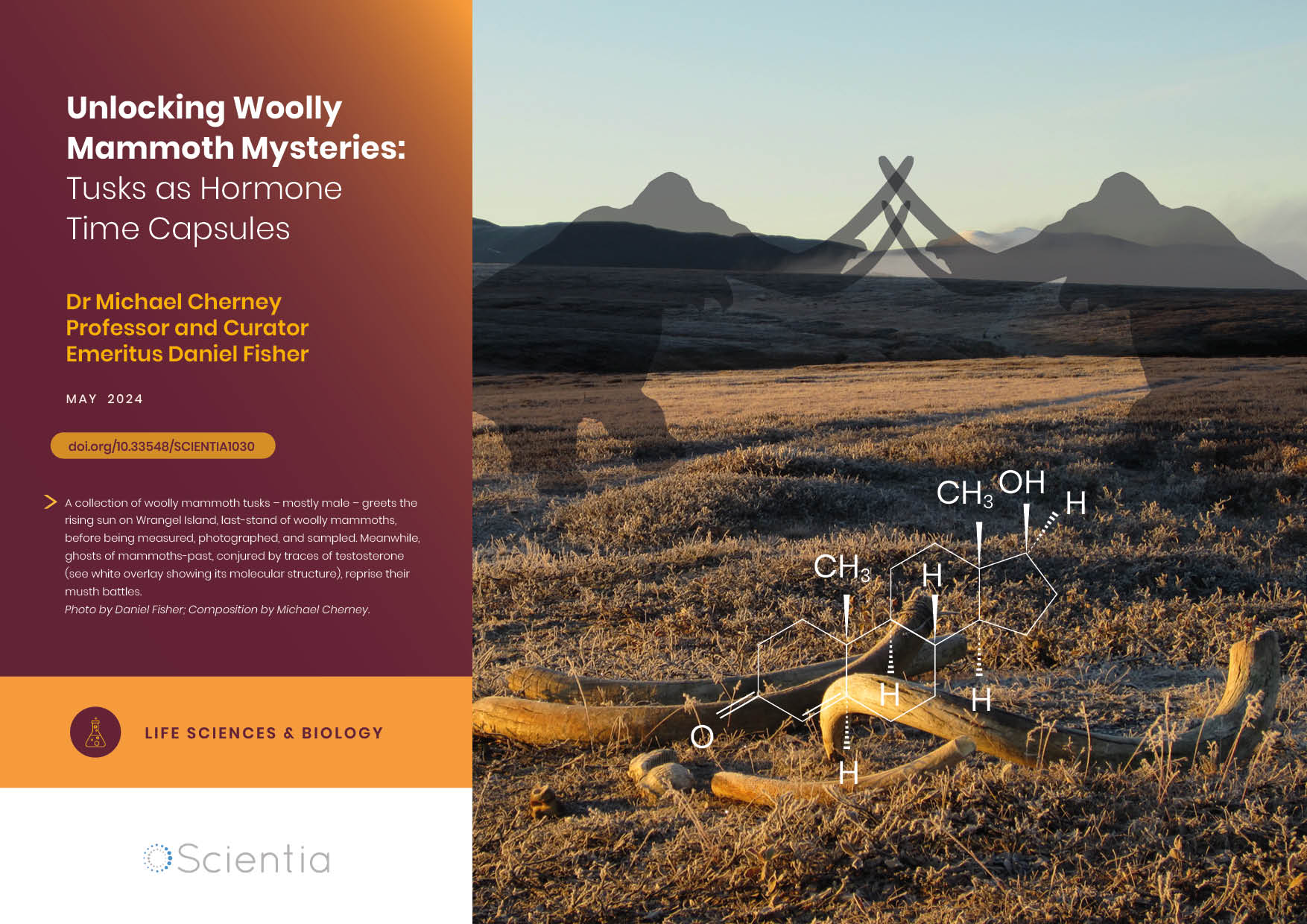
Dr Michael Cherney – Professor Daniel Fisher | Unlocking Woolly Mammoth Mysteries: Tusks as Hormone Time Capsules
The impressive tusks found on proboscideans (the order of mammals that includes elephants, woolly mammoths, and mastodons) are like time capsules, preserving detailed records of their bearers’ lives in the form of growth layers and chemical traces. Frozen in time for thousands of years, these layers can unlock secrets about the lives of long-extinct relatives of modern elephants. Dr Michael Cherney and Professor Daniel Fisher from the University of Michigan used innovative techniques to extract and analyse steroid hormones preserved in woolly mammoth tusks. This ground-breaking work opens new avenues for exploring the biology and behaviour of extinct species.