
Dr Kara Pratt | From Neurons to Behaviour: Exciting Insights from the Xenopus Tadpole
Understanding how neurons come together and form circuits in the brain is crucial to understanding how the brain works. Dr Kara Pratt and her team at the University of Wyoming are uncovering the mysteries behind the formation of neural circuitry and the ability of neurons to self-organise into highly refined networks. An incredibly elegant series of experiments using the larva stage of frogs has progressed insight into this fundamental phenomenon of neuroscience, specifically in the visual system.
Neurons: The Puzzle Pieces of the Brain
Neurons are the basic building blocks of the nervous system in the brain. They self-organise into circuits that allow correct brain functioning and information processing that ranges from basic sensory perception to complex cognitive processes. Imagine you have puzzle pieces, and you want to understand how they come together and form the puzzle image. In the world of neuroscience, this is a pivotal challenge, in which neurons are puzzle pieces that self-organise to form neural circuits. This question is crucial for a better understanding of the development of the brain and related processes.
Many psychiatric and neurological disorders are linked to problems in neural circuitry. Therefore, understanding neuronal assembly into neural circuitry is crucial not only for understanding how a healthy brain develops and functions throughout life but also for deeper insights when something goes wrong.
The Tiny Brain Wonders of Xenopus Tadpoles
There are various methodologies that scientists use to delve into the intricate world of neural circuitry and the neuronal ability to self-assemble. One captivating model to unveil these phenomena is the Xenopus tadpole, which is the larva stage of a frog. It provides an important scientific model allowing us to unravel the intricacies of how tadpoles transmit information from their eyes to their brains, and how these connections help them react to objects and events.
Leading the charge in this fascinating area are Dr Kara Pratt and her dedicated team at the University of Wyoming. They are especially interested in advancing the knowledge of the molecular pathways that underlie the process of neuronal self-organisation into highly refined, functionally distinct networks. More specifically, their work elucidates how different components of the visual system are involved in processing different aspects of the visual scene, and how this visual information consequently impacts behaviour.
These vital insights are gleaned from recording the electrical activity of individual neurons in the developing visual systems of Xenopus tadpoles and, alongside this, studying the visual system at the behavioural level. This combined approach provides a particularly powerful model with which to understand how neural circuits form and give rise to behaviours.

Studying Visually Guided Behaviours
As a postdoctoral researcher in Dr Carlos Aizenman’s laboratory at Brown University, Dr Pratt was part of the team that identified an exciting new behaviour in tadpoles. A novel behavioural assay known as a ‘moving dot test’ allowed the team to characterise visual avoidance behaviour in the laboratory, that is, when tadpoles swim away from specific visual stimuli. In the wild, this often occurs in potentially dangerous situations and evolved as a survival mechanism. The moving dot test takes advantage of the observation that tadpoles will usually dodge moving dots projected onto the floor of their test dish. These moving dots serve as visual cues that tadpoles respond to by swimming away. Dr Pratt is now working to explain how the visual system in tadpoles transforms such visual (i.e., sensory) input into motor output (i.e., behaviour).
From Seeing to Avoiding to Attraction
In a state-of-the-art review, Dr Pratt’s team summarised the existing literature on the topic of visual avoidance behaviour, pointing to the critical role of the tadpole retinotectal circuit in visual avoidance behaviour. This circuit is made up of special cells in their eyes that send the message to the optic tectum, the part of the amphibian brain that processes visual stimuli. A properly formed and refined retinotectal projection is essential for visual-avoidance behaviour, and it is thus unsurprising that it develops quickly in young tadpoles, with immature connections between the eye and tectum forming within days of fertilisation, maximising their chances of survival.
With the help of research students in her team, Dr Pratt set up a novel experimental design to answer two key questions. The first one was whether there were any other visually guided behaviours in tadpoles. If so, the second question was to find out whether those were also related to the retinotectal projection system – or some other component of the circuit.
In an important departure from previous research, the team investigated whether there was a visual stimulus that the tadpoles would swim towards (i.e., a stimulus that they would be attracted to rather than one they would avoid). They achieved this using a novel visual preference paradigm to measure the long-term preferences of freely swimming tadpoles.
Tadpoles Prefer the Colour Green (But How?)
This novel experiment showed, for the first time, that tadpoles prefer the colour green – in other words, they spent most of their time in the green area of the test dish. They preferred green when green was pitted against light, and they preferred green when green was pitted against dark.
Bearing in mind that the entire visual system comprises many distinct circuits, Dr Pratt was keen to identify which circuit(s) of the visual system are required for this behaviour in the tadpoles to take place. To this end, the team surgically removed different parts of the circuit and repeated the experiments. This revealed that the midbrain tegmentum (part of the visual processing centre) was critical, whereas the optic tectum, another visual processing centre, was not required for the tadpoles to display the innate preference for green. Notably, in humans, Dr Pratt notes that this brain area is also involved in different movement and motivation functions.
But how is it that tadpoles prefer the colour green? In other words, how is this preference for green manifested? The team determined that the preference for green is manifested by tadpoles slowing down, oftentimes even stalling, when they are in the green region of the test dish. Thus, for tadpoles, green means ‘stop!’.
Dr Pratt suggests that this could be caused by the neurotransmitter serotonin (a brain molecule that regulates various physiological and psychological processes in the body). Her premise is based on research from Professor Nick Spitzer’s laboratory, revealing that increased levels of serotonin result in shorter swimming bouts. Dr Pratt and her team suspected that changes in serotonin levels may influence tadpoles’ preference for the colour green, too.
To test this, the researchers exposed tadpoles to selective serotonin reuptake inhibitors (SSRIs) for 24 hours. These drugs are used to control levels of serotonin in depression, for example. To Dr Pratt’s surprise, the SSRIs did not increase the existing preference for green. Regardless of the experimental manipulation, tadpoles consistently preferred the brightest part of the dish. This gave the researchers a new and intriguing idea – the SSRIs shifted the preference from colour-based to luminance-based.

Credit: Harley Yerdon
Serotonin’s Role in Tadpoles’ Visual Preference
These latest findings and possibilities spurred the team to explore the visual preference of tadpoles further. In the next series of experiments, the team switched to a simpler design of ‘light versus dark’ to test only the preference for luminance. The results were fascinating! The study showed that the tadpoles’ preference for luminance peaks at around 4 pm and is lowest at night. When the team used the SSRIs to enhance levels of serotonin transmission, the preference for light increased significantly. However, when serotonin levels were decreased by exposing tadpoles to an inhibitor of tryptophan hydroxylase (an enzyme that synthesises serotonin), the preference for light decreased, too. This strongly suggests that serotonin, the naturally occurring chemical in the brain, creates the preference for light over dark in Xenopus tadpoles.
Putting Together the Pieces
Dr Pratt has advanced insights into the visual preferences of tadpoles, and how these preferences take place. First, her work shows that normally, tadpoles display a modest yet significant preference for green over light, and also a preference for light over dark. Second, she has shown that artificially enhancing the level of serotonin transmission leads to an abnormally strong preference for light. Finally, we now know that inhibiting serotonin levels in tadpoles significantly reduces the preference for light.
These patterns likely reflect survival mechanisms. Dr Pratt and the team theorise that spending time in well-lit regions during the day may be important in order to find food or to dwell near sun-warmed regions of water. Similarly, it may be important for them to spend time near green plants that are a source of food and safety. This preference, which is neither too strong nor too weak, may be optimal for their survival according to so-called predator-prey interactions theories. These models suggest that being too predictable in behaviour could make tadpoles more vulnerable to predators.
Having established the importance of the maintenance and timely release of the optimal levels of serotonin during the day, Dr Pratt’s work highlights that naturally occurring brain chemicals such as serotonin may play a crucial role in shaping how neural circuits form and function. She is now investigating the underlying neural circuits that give rise to these visual preference behaviours and, importantly, how serotonin modulates these circuits.
SHARE
DOWNLOAD E-BOOK

REFERENCE
https://doi.org/10.33548/SCIENTIA1013
MEET THE RESEARCHER
Dr Kara Geo Pratt
Department of Zoology and Physiology
and Graduate Program in Neuroscience
Laramie, WY
USA
Dr Kara Pratt received her PhD in Neuroscience from Brandeis University in 2004. After undertaking several postdoctoral positions at Brown University, the University of Washington, and Massachusetts General Hospital, she started her own laboratory at the University of Wyoming. She has worked as an Assistant Professor in the Department of Zoology and Physiology since 2011 and was promoted to Associate Professor in 2017. Dr Pratt is specifically interested in researching the molecular pathways underlying the developing visual systems of the Xenopus tadpole. She has been supported by prestigious funding and has published over 28 peer-reviewed journal articles to date. On top of her research endeavours, she teaches courses in neurodevelopmental and developmental biology.
CONTACT
E: Kpratt4@uwyo.edu
W: www.uwyo.edu/zoology/people/pratt.html
www.uwyo.edu/neuroscience/people/fac_landing/pratt.html
FUNDING
National Science Foundation
National Institutes of Health
FURTHER READING
JR Bruno, UG Udoh, LG Landen, et al., A circadian-dependent preference for light displayed by Xenopus tadpoles is modulated by serotonin, iScience, 2022, 25(11), 105375. DOI: https://doi.org/10.1016/j.isci.2022.105375
JE Hunt, JR Bruno, KG Pratt, An innate color preference displayed by Xenopus tadpoles is persistent and requires the tegmentum, Frontiers in Behavioral Neuroscience, 2020, 14, 71. DOI: https://doi.org/10.3389/fnbeh.2020.00071
Z Liu, AS Hamodi, KG Pratt, Early development and function of the Xenopus tadpole retinotectal circuit, Current Opinion in Neurobiology, 2016, 41, 17–23. DOI: https://doi.org/10.1016/j.conb.2016.07.002
![]()
REPUBLISH OUR ARTICLES
We encourage all formats of sharing and republishing of our articles. Whether you want to host on your website, publication or blog, we welcome this. Find out more
Creative Commons Licence (CC BY 4.0)
This work is licensed under a Creative Commons Attribution 4.0 International License. 
What does this mean?
Share: You can copy and redistribute the material in any medium or format
Adapt: You can change, and build upon the material for any purpose, even commercially.
Credit: You must give appropriate credit, provide a link to the license, and indicate if changes were made.
SUBSCRIBE NOW
Follow Us
MORE ARTICLES YOU MAY LIKE

Professor Ken M Levy | The Boundaries of Free Will and Responsibility: From Academic Debate to the Real World
For almost thirty years, Professor Ken M Levy of Louisiana State University Law School has been thinking and writing about free will and responsibility. In several articles and his recent book, Free Will, Responsibility, and Crime: An Introduction (Routledge 2020), Professor Levy discusses a wide range of subjects, including the myth of the ‘self-made man’, whether psychopaths are culpable for their crimes, and the increasingly popular but highly controversial theory of responsibility scepticism. Professor Levy’s research has profound implications for law, ethics, and society.

Dr Michael Hoffmann | Mirror Neurons as a Key to Stroke Rehabilitation
Mirror neurons are specialised brain cells that underpin our capacity to learn and understand a myriad of behaviours. Dr Michael Hoffmann, from the University of Central Florida and the Roskamp Institute in Florida, has unravelled the profound implications of these brain cells. Beyond their role in cognition, mirror neurons could play a major role in patient rehabilitation, particularly in the context of stroke recovery.
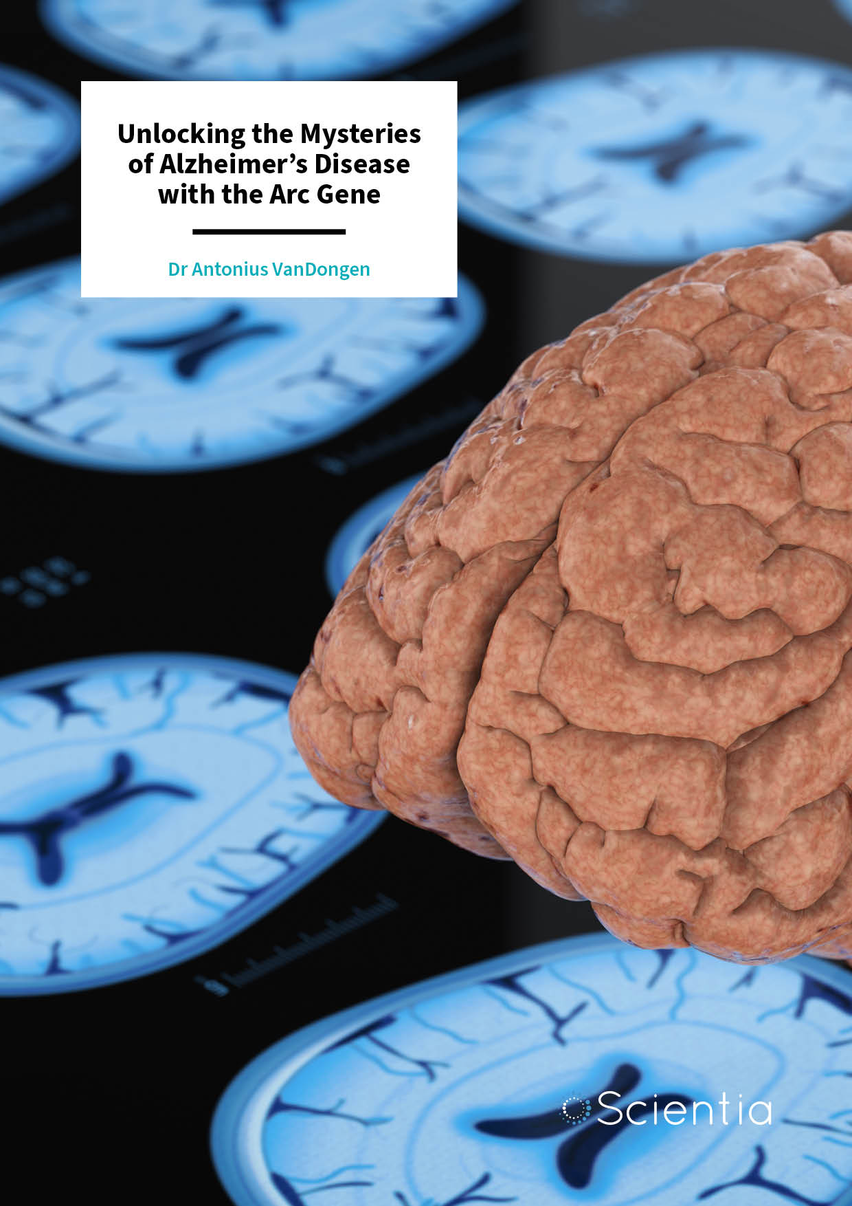
Dr Antonius VanDongen | Unlocking the Mysteries of Alzheimer’s Disease with the Arc Gene
Our vulnerability to developing diseases and conditions depends upon a complex interaction between our genes, lifestyle, and environmental factors. Alzheimer’s disease is no exception to this, and sadly, it remains without a cure. Dr Antonius VanDongen and his team from Duke University are studying the mechanisms underlying learning and memory, specifically focusing on the activity-regulated cytoskeletal memory gene Arc. Their work is driving forward our understanding of the memory problems that characterise Alzheimer’s disease.

Dr Audrey Nath | Neurological Disorders in Prisoners: A Neglected but Complex Problem
Incarceration presents a unique set of challenges for the health of individuals, particularly when it comes to neurological conditions. In a comprehensive review, Dr Audrey Nath and Samuel Han have delved into the often-overlooked realm of neurological health. From learning disabilities to epilepsy, sleep disorders, infectious diseases, nutritional deficiencies, toxicology-related issues, and traumatic brain injuries, their review sheds important light on the complex landscape of neurological health within the prison system.