
Dr Linda Hammerich | Revolutionising Immune Monitoring with Flow Cytometry
Understanding the individual immune response is key to diagnosing and treating a range of diseases. One way of characterising immune cells is through flow cytometry, where cells are tagged with fluorescent markers known as fluorochromes. Detectors use these markers to understand the different physical and chemical features of the individual cells and the overall immune cell population. Dr Linda Hammerich and a team from Charité-Universitätsmedizin in Germany have optimised this technique to investigate up to 31 different cells or markers from one blood test using currently available technology.
The Immune System and Flow Cytometry
The immune system is a complex network of cells, proteins, and molecules that defend the body against infections, disease, and other foreign invaders. Its primary role is recognising and neutralising pathogens such as bacteria, viruses, fungi, and parasites. Immune cells, such as white blood cells like lymphocytes, macrophages, and neutrophils, are key players in this defence mechanism. Beyond fighting infections, the immune system plays a vital role in identifying and eliminating cancer cells. It also helps in wound healing and tissue repair.
However, an overactive immune response can lead to autoimmune diseases, where the body mistakenly attacks its own tissues, and allergic reactions, where harmless substances trigger an immune response. Thus, the immune system’s varied functions are essential for maintaining overall health and protecting the body from a wide range of threats. Understanding a patient’s immune response is key to providing appropriate care, particularly in the current age of personalised medicine.
Flow cytometry is a powerful laboratory technique used to analyse the physical and chemical characteristics of cells or particles suspended in a fluid. It operates on the principle of passing these cells through a laser beam, one cell at a time, and measuring the light they scatter and the fluorescence they emit. The process begins with cells being stained with fluorescent dyes known as fluorochromes that bind to specific cellular components. Detectors capture the scattered light and fluorescence that is emitted as these stained cells pass across the laser.
Forward scatter (FSC) provides information about cell size, while side scatter (SSC) gives insights into cell complexity or granularity. Fluorescent markers reveal specific features, such as surface proteins, DNA content, or other cellular functions.
Flow cytometry is widely used in healthcare to diagnose and monitor various diseases. In immunology, it’s essential for identifying and counting different types of immune cells, which is crucial for managing conditions like HIV and autoimmune disorders. In oncology, flow cytometry helps diagnose blood cancers such as leukaemia and lymphoma by identifying abnormal cell populations and monitoring the immune cell population.
Full Spectrum Flow Cytometry
As our understanding of the immune system and its role in different diseases has become more advanced, it has become increasingly valuable and important for clinicians to analyse as many immune cells and markers from one blood test as possible. Traditional flow cytometry has been useful at answering important but simple questions, such as how many of one type of cell there are in someone’s blood, but it has not been able to answer more detailed questions.
Full-spectrum flow cytometry is an advanced version of flow cytometry. Unlike the traditional method, which uses a few specific filters to detect different colours of light, this method uses many detectors to capture the entire range of light emitted by the fluorochromes attached to the cells. This allows scientists to simultaneously analyse many different markers on a single cell, giving them a much more detailed picture of its characteristics.

Gaining the Maximum Benefit from Existing Technology
To help understand the potential for full-spectrum flow cytometry to be used on the traditional flow cytometry machines currently found in healthcare and research labs, Dr Linda Hammerich and her team of scientists at Charité-Universitätsmedizin in Germany have been conducting research to calculate the maximum number of cell markers that can be detected using standard equipment and fluorochromes.
The team conducted careful planning and experiments using real cells to design a panel of 31 fluorochromes to analyse different aspects of immune cells from human blood tests. They worked with blood donated by healthy volunteers and antibody/fluorochrome combinations that had already been tested and optimised in their lab on traditional flow cytometers. They followed guidelines from the flow cytometer machine manufacturer (Cytek Biosciences) for typical 3-laser machines to select an initial set of 23 fluorochromes.
Then, they gradually added more dyes, ensuring each new one had a distinct colour profile from those already included. For immune markers that are usually low in healthy people, they swapped them out for markers with higher levels to ensure all 31 markers could be clearly identified. They checked if the antibodies worked as well on the Cytek AuroraTM spectral flow cytometer as they did on conventional machines found in hospitals and research laboratories. This was the case when they were used at the same concentration. If an antibody did not show clear results, they adjusted its concentration using a process called titration. They performed controls to find the optimal amount for clear detection for each fluorochrome.
The team’s work had very exciting results. They have produced a panel that can identify all the major immune cell types that would be found in a human blood test, including types of cells that are only found at very low frequencies. The method does not require any custom setup and utilises commercially available fluorochromes, making it especially valuable to healthcare systems with financial limitations worldwide. The panel can also act as a starting point or a model for other researchers and healthcare professionals to create similar multi-panel markers studying immune cells, as the panel can be tailored to different research or clinical needs.
The team are especially excited by the fact that their detection panel only utilised 29 of the 38 detectors available on the machine due to overlapping fluorescence, meaning there is potential to build from their work and develop even more detailed panels as biotechnology companies develop novel fluorochromes.
SHARE
DOWNLOAD E-BOOK

REFERENCE
https://doi.org/10.33548/SCIENTIA1124
MEET THE RESEARCHER

Dr Linda Hammerich
Department of Gastroenterology, Charité-Universitätsmedizin, Berlin, Germany
Dr Linda Hammerich gained her PhD in liver inflammation from RWTH Aachen University in Germany. She then spent five years as a postdoctoral researcher at the Tisch Cancer Institute at Icahn School of Medicine at Mount Sinai in New York City, USA. She now leads a research group at Charité-Universitätsmedizin in Berlin, Germany, where she specialises in tumour immunology and immunotherapy in gastrointestinal cancers. She is leading research on developing vaccines for liver cancer. She is also committed to reducing the use of animals in biomedical research by optimising traditional techniques such as flow cytometry to gain the most information from a single sample. Alongside her research, she supports MSc, PhD, and medical students with their research projects. She has been invited to share her expertise by speaking at medical and immunological conferences worldwide.
CONTACT
X: @Flt3Linda
FUNDING
Charité 3R│Replace – Reduce – Refine
FURTHER READING
Y Shevchenko, I Lurje, F Tacke, L Hammerich, Fluorochrome-dependent specific changes in spectral profiles using different compensation beads or primary cells in full spectrum cytometry, Cytometry A, 2024, 105(6), 458–463. DOI: https://doi.org/10.1002/cyto.a.24836
L Hammerich, Y Shevchenko, J Knorr, et al., Resolving 31 colors on a standard 3-laser full spectrum flow cytometer for immune monitoring of human blood samples, Clinical Cytometry, 2023, 104(5), 367–373. DOI: https://doi.org/10.1002/cyto.b.22126
REPUBLISH OUR ARTICLES
We encourage all formats of sharing and republishing of our articles. Whether you want to host on your website, publication or blog, we welcome this. Find out more
Creative Commons Licence (CC BY 4.0)
This work is licensed under a Creative Commons Attribution 4.0 International License. 
What does this mean?
Share: You can copy and redistribute the material in any medium or format
Adapt: You can change, and build upon the material for any purpose, even commercially.
Credit: You must give appropriate credit, provide a link to the license, and indicate if changes were made.
SUBSCRIBE NOW
Follow Us
MORE ARTICLES YOU MAY LIKE

Professor James Calvin | Innovation in Heart Failure Care: Comparing Personal Coaching with Digital Support
Heart failure remains one of the most challenging conditions facing healthcare systems today, with hundreds of thousands of new cases diagnosed annually. Professor James Calvin from Western University’s Schulich School of Medicine and Dentistry has led groundbreaking research comparing two innovative approaches to supporting patients: personal health coaches and smartphone reminders. His team’s findings suggest that combining human support with digital technology could transform how we help patients manage this complex condition.

Dr Hermann Salmhofer | Minimising the Damage Caused by Systemic Viral Infections
The mechanisms via which viral diseases infect and progress within the human body have become the subject of intense scrutiny since the emergence of the serious respiratory condition COVID-19, although many other viruses remain woefully under-researched. Recently, Dr Hermann Salmhofer and colleagues at the Paracelsus Medical University in Salzburg, Austria, have described the successful treatment of a harmful systemic virus affecting the kidneys, and suggest a possible new treatment target to mitigate the progression of the disease and prevent the development of permanent organ damage. Their findings, combined with broader research on viral infections, highlight the critical importance of both preventive measures and targeted treatments in managing viral diseases.

Revealing the Intricate Links Between Metabolism and Reproduction
The brain plays a vital role in controlling reproductive functions. It helps to maintain a delicate balance of hormones, all of which can be affected by the metabolism. Investigating the impact of the metabolism on reproductive development and function is critical to a better understanding of health and diseases. Professor Carol Fuzeti Elias and Dr Cristina Sáenz de Miera Patín from the University of Michigan in the USA, carry out groundbreaking research in neuroscience, exploring the molecular and neural mechanisms at play.
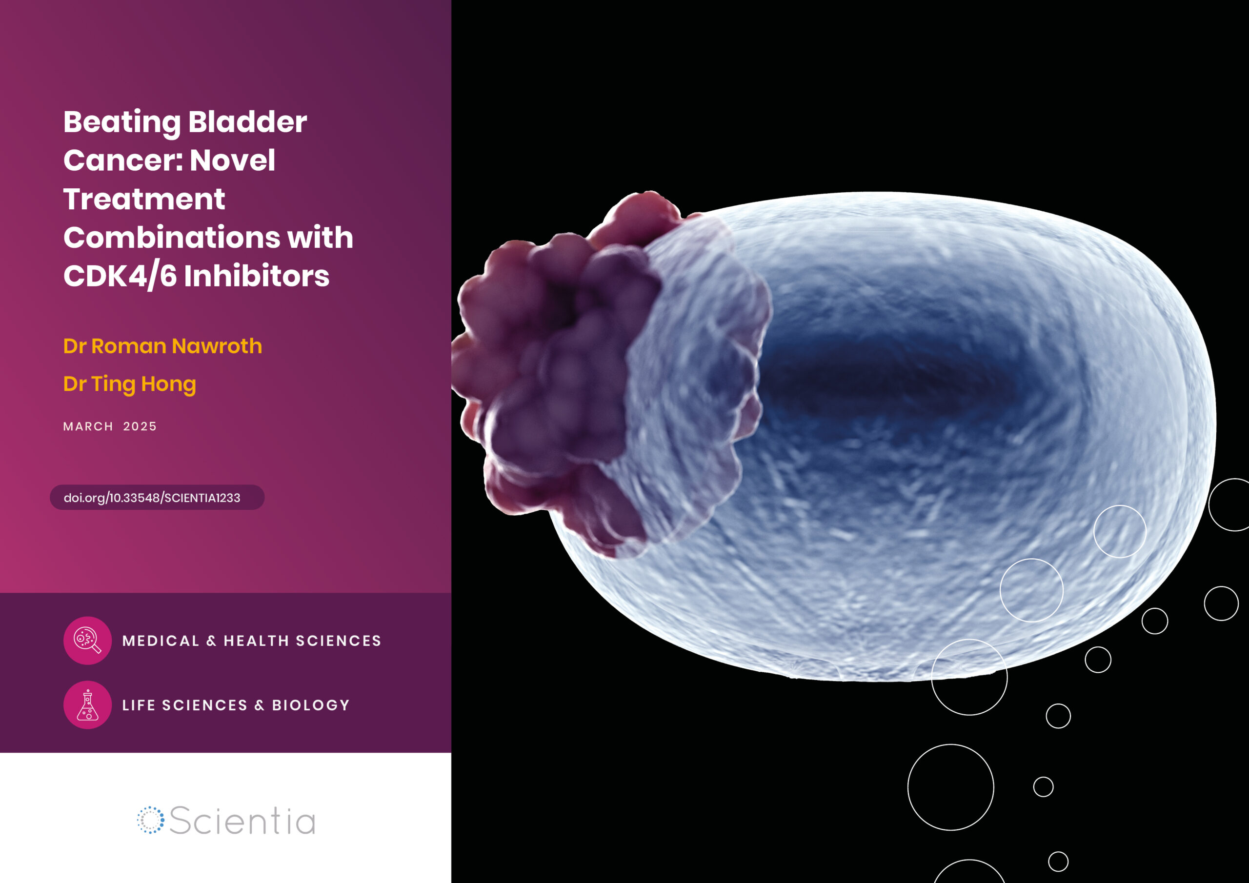
Beating Bladder Cancer: Novel Treatment Combinations with CDK4/6 Inhibitors
Cancer is one of the leading causes of death around the world. Research into this disease is vital to the development of new treatments, bringing fresh hopes to those affected by this potentially devastating diagnosis. Dr Roman Nawroth and Dr Ting Hong carry out their ground-breaking research at the Technical University of Munich in Germany. They focus their efforts on novel approaches to fight bladder cancer, exploring the use of CDK4/6 inhibitors.