
Dr Martin Schwarz – Mapping the Brain’s Neuronal Networks to Understand Behaviour
The human brain is wonderfully complex. Billions of neuron cells connect in unique ways to create networks that determine each individual’s brain function and consequent behaviour. Given the expanse and complexity of these networks, it is not surprising that they are not yet fully mapped out or understood. Bringing innovative new and exciting ideas to this field of neurobiology is Dr Martin Schwarz from the Institute for Experimental Epileptology and Cognition Research and the Life & Brain Center at the University of Bonn Medical Center in Germany.
Neurons: The ‘Talking’ Cells in the Brain
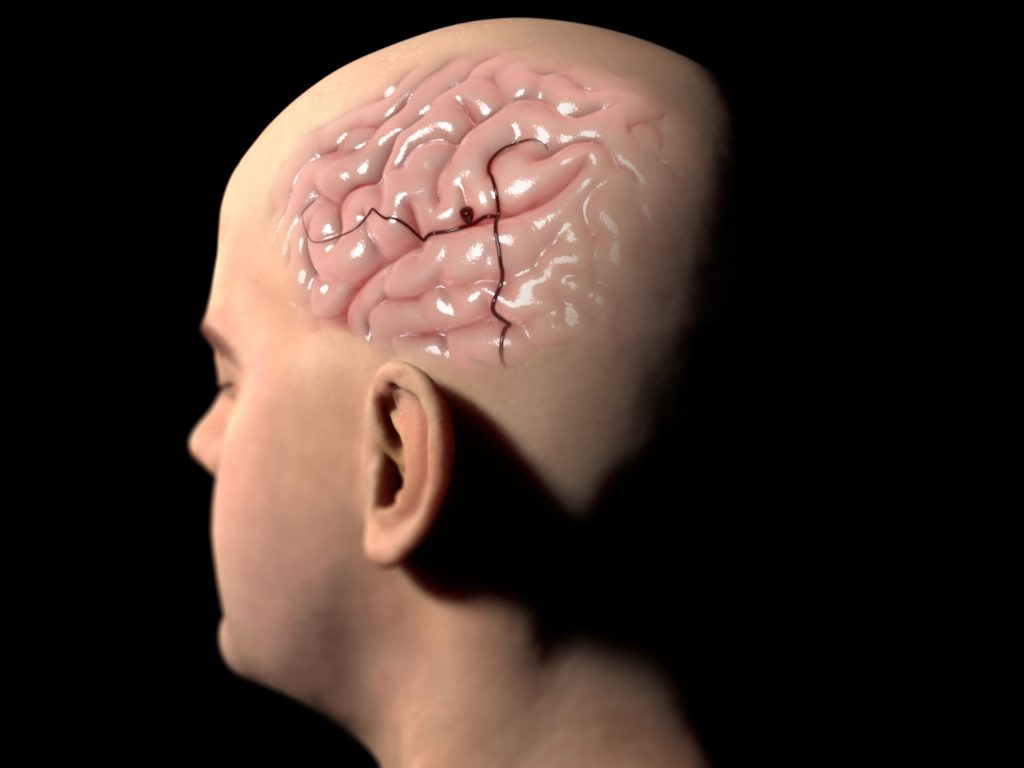
Our brains are made up of a staggering 86 billion neuron cells connecting and working together to keep our minds alive. Neurons are one of the two types of cells in the nervous system, the others are glial cells. They have a cell body that holds the nucleus with its genetic material and controls the activities of the cell. From this body, extensions called dendrites branch out to talk with adjacent axons, the long tail of the cell that transmits messages.
These messages are sent and received in the form of chemicals called neurotransmitters, such as dopamine, serotonin and adrenaline, among hundreds of others. Neurotransmitters are sent across the tiny spaces, known as synapses, between the dendrites and axons to deliver information across the brain from neuron to neuron. Neurons typically serve specific functions and are therefore categorised according to their main function such as sensory, motor or interneurons. Sensory neurons carry information from the sensory organs, like the nose and eyes, to the brain whilst motor neurons control voluntary muscle movement like speaking by sending messages from the brain to the muscles.
Because there are so many different types of neurons, each with their own set of attributes and functions, and because each person has a distinctive network of them, the minds of humans are unique. The number and types of neuronal connections in the brain determine how a person thinks, feels and behaves, making them an essential part of the body. However, due to the complex and extensive nature of these neuronal networks, our understanding of how they all fit and work together is not yet complete.
As a result, there is also a gap in our knowledge in the way in which these circuits impact behaviour. Working to fill this knowledge gap is Dr Martin Schwarz from the Institute for Experimental Epileptology and Cognition Research and the Life & Brain Center at the University of Bonn Medical Center in Germany. Dr Schwarz explains that ‘a prerequisite to understanding these “brain algorithms” is to understand a circuits’ hardware – in other words: who is synapsing with whom, how often and how does this concerted activity then result in a specific behaviour.’
To achieve these insights, Dr Schwarz is developing, employing and combining novel methods to determine how neuronal structure and function are linked to complex behaviour. Importantly, the success of this requires active collaboration with researchers from other disciplines, providing testimony to the power of inter- and multi-disciplinary research in driving scientific advancement.

Mapping and Manipulating Neuronal Networks with Artificial Intelligence
Dr Schwarz and his team develop and utilise clever technologies to help map and characterise the neuronal circuits involved in the continuous processing of information. One such technology is an artificial intelligence system called DeepLabStream that recognises the behaviour of mice and facilitates the labelling of the neuronal networks that are actively involved in that behaviour.
In an experimental setup using DeepLabStream, Dr Schwarz and his co-worker Jens Schweihoff placed mice in a box and allowed them to move freely. Whenever the mouse looked in a specific direction, the behaviour was detected by DeepLabStream and the involved neuronal circuits were labelled for subsequent analysis.
For this, Dr Schwarz and his team made use of a set of proteins brought into the brain, called Cal-Light, that can be used to label neurons that are active during specific durations by shining a light into the brain. In combination with DeepLabStream, the scientists could use the system to capture neuronal networks during selected behaviour. This effectively enabled them to determine which neuronal networks were active during specific behaviours in real-time.
This technique also allows researchers to manipulate neuronal activity during any type of behaviour – such as learning and memory – in a short-term manner. This is an alternative to longer-lasting and behaviour-altering manipulations such as creating brain lesions which often result in long-term changes in the brain that make it difficult to relate behaviour back to neuronal networks.
These are exciting firsts for the field and Dr Schwarz believes they have promising implications. Thanks to the DeepLabStream system’s improved accuracy, Cal-Light can now be utilised effectively in a broader range of experiments and reduce variability across the board. The software is open source so scientists across the globe can contribute to improving our understanding of neurobiology.
‘Our long-term goal is to anatomically dissect and functionally characterise neuronal network components to better understand how erroneous neuronal wiring finally translates into specific behavioural deficits.’

“This experimental strategy has the potential to untangle
previously unknown causal relationships between brain
activity and behaviour” Jens Schweihof
Visualising Transplant Connections with the Rabies Virus
As part of another study, Dr Schwarz and his co-workers visualised the underlying biology of the brain in a different way. When a person sustains a brain injury or develops a brain disease, their neuronal cells may die and thus be lost. To combat this issue, researchers, including Dr Schwarz, are innovating methods of replacing these cells through transplantation. For example, in the neurodegenerative disorder Parkinson’s disease, it would be extremely useful to reintroduce the neurons that produce and transmit the dopamine that is lost.
However, until now, it has been difficult to ascertain whether transplanted neurons actually reconnect with those around them to produce an operational neuronal network and restore normal function. Dr Schwarz’s team, together with the team of Prof Oliver Brüstle, used an unusual source in their quest to identify a new technique for seeing implanted cells within the brain in high resolution.
The researchers used genetically engineered rabies viruses that were no longer harmful but still travelled via their usual method of spreading backwards through the nervous system, across the synapses. Attached to each virus was a fluorescent protein that allowed the team to observe a green glow under a microscope when a transplanted neuron was connected to an existing one.
In the study, Dr Schwarz and his co-workers transplanted human stem cells into two brain regions of mice, which matured into neurons. They were then infected with the modified rabies virus and the brains underwent a procedure that made them as transparent as glass so any fluorescence can be seen throughout the entire brain. The relevant section of the brain is scanned with a light-sheet microscope to create a three-dimensional map of the connected transplant and recipient neurons.
Using this exciting technique, the team was able to reveal that their transplants were incorporated into precisely the right neuronal circuits in the target regions. This is an exciting result because it holds the potential to guide clinical studies in the future.
Light Sheet Fluorescence Expansion Microscopy
A significant portion of Dr Schwarz’s work aims to improve current technologies to expand the boundaries of research. The previously mentioned Cal-Light technique needs a type of light-sheet fluorescence microscopy – one of the technologies that Dr Schwarz’s group studies and improves. For example, his team together with the team of Prof Ulrich Kubitscheck developed a light-sheet fluorescence microscopy method that allowed the evaluation of large sections of brain tissue at very high resolutions previously unseen.
To achieve this, they used a technique known as tissue expansion, in which the brain sample is placed within an expandable polymer and evenly expanded to a multitude of its original size. When combined with light-sheet fluorescence microscopy, it allows even the tiny synapses between the neurons to be seen fluorescing under a microscope. For this reason, this method is known as light-sheet fluorescence expansion microscopy. These novel ways of visualising brain tissue and the neuronal circuits within will help researchers to continue to build up a map of these complex networks and link them to behaviour.

The Power of Smell
Of particular interest to Dr Schwarz is the behavioural response elicited by smells. At the bottom of the brain is a bilateral structure called the horizontal diagonal band (HDB). Although it plays an important role in the olfactory (habituation) system, the HDB does not directly receive olfactory input. Instead, it indirectly recognises when odour information is incoming and regulates odour sensation within a primary olfactory processing region called the olfactory bulb.
Dr Schwarz and his team investigated this neuronal circuit in relation to olfactory habituation – the behaviour of repetitive, irrelevant odour stimuli being ignored to allow more processing of significant odour input. His studies showed that when the HDB is inhibited, olfactory habituation and the ability to distinguish different odours are completely prevented. This demonstrates a causal link between the two regions and the research team hypothesised that the neuronal circuits that actively control olfactory habituation and discrimination reside within the HDB. Moving forward, Dr Schwarz and his co-workers aim to map these networks using the Cal-Light system and determine the functional circuits of distinct odours to dissect odour specific neuronal ensembles.
Speaking about the entirety of his work to date, Dr Schwarz explains, ‘Our long-term goal is to anatomically dissect and functionally characterise neuronal network components to better understand how erroneous neuronal wiring finally translates into specific behavioural deficits.’
Dr Schwarz has already pushed forwards our understanding of neuronal networks. He concludes by noting that future technical developments in this field will certainly require the utilisation of artificial intelligence-based systems to facilitate a more precise and less error-prone analysis of behavioural algorithms and their neuronal implementation in health and disease.
SHARE
DOWNLOAD E-BOOK
REFERENCE
https://doi.org/10.33548/SCIENTIA781
MEET THE RESEARCHER

Dr Martin K. Schwarz
Institute for Experimental Epileptology and Cognition Research, and Life & Brain Center
University of Bonn Medical Center
Bonn
Germany
Dr Martin Schwarz received his doctor rerum naturalium (equivalent to a PhD) from the University of Vienna in cooperation with the Max Planck Institute for Biophysical Chemistry in Göttingen in Germany. Over the course of his career, Dr Schwarz has held a range of positions at prestigious German institutions (Max-Planck Institute of Medical Research in Heidelberg) and currently serves as the Principal Investigator of the Functional Neuroconnectomics group at the Institute of Experimental Epileptology and Cognition Research and is also affiliated with the Life & Brain Center at the University of Bonn Medical Center in Germany. His work focuses on the development of cutting-edge methods to facilitate the identification and characterisation of neuronal networks within the brain to better understand the correlation between neuronal activity and behaviour.
CONTACT
KEY COLLABORATORS
Prof Dr Heinz Beck, University of Bonn, Germany
Prof Dr Ulrich Kubitscheck, University of Bonn, Germany
Prof Dr Oliver Brüstle, University of Bonn, Germany
Prof Dr Martin Fuhrmann, German Center for Neurodegenerative Diseases, Germany
Prof Dr Stefan Remy, Leibniz-Institut für Neurobiologie, Germany
FUNDING
Deutsche Forschungsgemeinschaft (DFG)
National Institutes of Health (NIH)
FURTHER READING
JF Schweihoff, M Loshakov, I Pavlova, et al., DeepLabStream enables closed-loop behavioral experiments using deep learning-based markerless, real-time posture detection, Communications Biology, 2021, 4, 130.
J Bürgers, I Pavlova, JE Rodriguez-Gatica, et al., Light-sheet fluorescence expansion microscopy: fast mapping of neural circuits at super resolution, Neurophotonics, 2019, 6(1), 015005.
J Doerr, MK Schwarz, D Wiedermann, et al., Whole-brain 3D mapping of human neural transplant innervation, Nature Communications, 2017, 8, 14162.
MK Schwarz, A Scherbarth, R Sprengel, et al., Fluorescent-Protein Stabilization and High-Resolution Imaging of Cleared, Intact Mouse Brains, PLoS One, 2015, 10(5), e0124650.
CJ Niedworok, I Schwarz, J Ledderose, et al., Charting Monosynaptic Connectivity Maps by Two-Color Light-Sheet Fluorescence Microscopy, Cell reports, 2012, 2(5), 1375–1386.
REPUBLISH OUR ARTICLES
We encourage all formats of sharing and republishing of our articles. Whether you want to host on your website, publication or blog, we welcome this. Find out more
Creative Commons Licence (CC BY 4.0)
This work is licensed under a Creative Commons Attribution 4.0 International License. 
What does this mean?
Share: You can copy and redistribute the material in any medium or format
Adapt: You can change, and build upon the material for any purpose, even commercially.
Credit: You must give appropriate credit, provide a link to the license, and indicate if changes were made.
SUBSCRIBE NOW
Follow Us
MORE ARTICLES YOU MAY LIKE

Professor Alan Templeton | When Fire Sparks Ecological Opportunity and Habitat Restoration
How far would you be willing to go to save an endangered species? Would you consider burning part of a forest as a solution? As unconventional as it may sound, conservationists sometimes resort to such measures to restore lost habitats. One remarkable example is the efforts to save eastern collared lizards – and indeed the entire biological community in which they live – in the Ozarks, spearheaded by American geneticist and statistician Professor Alan Templeton of Washington University in St Louis, USA.

Dr Toby Phesse | Revealing the Mysteries of Wnt Signalling: Novel Approaches to Beating Cancer
Cancer remains a leading cause of mortality worldwide, and the need for new, more effective treatments remains an urgent challenge. Dr Toby Phesse from Cardiff University in the UK focuses on the role of the Wnt receptor found on the surface of cells and its involvement with cell communication and cancer growth, bringing fresh hopes for new therapeutic options.
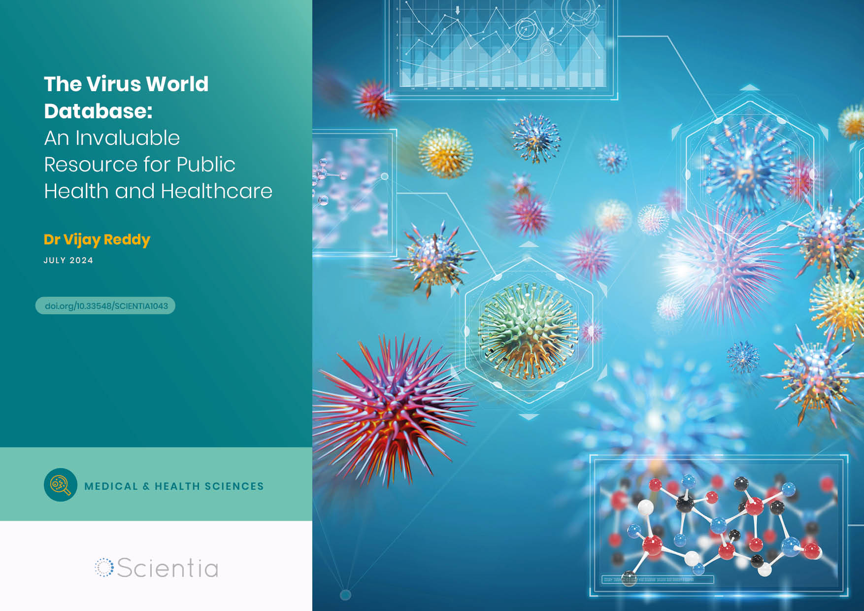
Dr Vijay Reddy | The Virus World Database: An Invaluable Resource for Public Health and Healthcare
Severe viral disease presents an ongoing challenge to the health of humankind. While unparalleled developments in science and technology are improving our understanding of such viruses, this information needs to be readily accessible to researchers to ensure continued progress in public health and healthcare. Dr Vijay Reddy and his colleagues at the Hormel Institute (University of Minnesota) developed the Virus World database, an invaluable resource that details the genome, structure, and host of practically every discovered virus to date.

Professor Ralf Herwig | Deciphering the Enigma of Vitamin D and the Immune System
Vitamin D has been studied as a treatment for a large number of diseases and conditions, from cancer to autism to COVID-19. However, its mode of action is not completely understood. Professor Ralf Herwig carries out his research at HG Pharma GmbH (Austria) and Ulster University (UK). His vital work explores the role of vitamin D in the body with a view to unlocking its potential as a treatment for a variety of health conditions involving the immune system.