
Dr Matthew Boisen – Understanding Lassa Virus
For many years, Dr Matthew Boisen, Director of Diagnostics Development at Zalgen Labs, has focussed on trying to understand Lassa fever. Part of the Viral Hemorrhagic Fever Consortium, his group’s objectives are threefold: first, to develop fast and accurate diagnostics for Lassa fever; second, to design new therapeutic approaches; and third, to create an effective vaccine providing long-term protection against this condition.
It is estimated that there are 59 million people at risk of contracting Lassa fever, primarily in West Africa, with 3.3 million potentially suffering severe morbidity after contracting the disease. Following an incubation period of 7 to 21 days, most people experience mild symptoms including fever, headache, and general weakness. However, about 1 in 5 patients can develop a more severe condition, with hypotension, facial oedema, and vomiting.
In a third of these cases, symptoms may progress to general internal bleeding, which is fatal for most patients. Death occurs 10–14 days after presentation of the initial symptoms, usually due to multi-organ failure. Pregnant women are particularly at risk, with mortality reaching 90% for mothers, and spontaneous abortion occurring in the majority of cases.
The Lassa virus is one of more than 30 known pathogens from the arenavirus family. The family has an Old World branch, including the Lassa virus as well as lymphocytic choriomeningitis virus, which causes fever and birth defects; and the haemorrhagic fever virus Lujo, first detected in 2008. New World viruses include the Machupo virus and Junín virus, responsible for the Bolivian and Argentinian haemorrhagic fever, respectively, as well as numerous other viruses, such as Sabía and Guanarito.
This virus infects more than 300,000 individuals per year, with Sierra Leone, Guinea, Liberia, and Nigeria reporting the highest incidences. Benin, Togo, Burkina Faso, and Mali have also reported cases over the past few years, and the virus has been exported from its normal geographic region, with cases identified recently in Germany, Sweden, the USA, and the United Kingdom.
Despite the seriousness of this condition, diagnosis can be difficult. The problem is that there are many other conditions which present similar symptoms, including malaria, typhoid fever, leptospirosis, and arbovirus diseases. To further complicate matters, the only available treatment – ribavirin – must be carried out during the first six days of illness to be effective. ‘Rapid diagnosis is imperative for proper patient management, which includes patient isolation, treatment with ribavirin when available, and supportive care such as the replacement of fluids and electrolytes,’ explains Dr Boisen.
Due to high mortality and ease of infection, the Lassa virus is at the centre of many attempts to accelerate the development of a new vaccine. Dr Boisen is a leading example, and his team at Zalgen Labs has been working hard to better understand the virus with the aim to develop simpler and more accurate diagnostic tools, as well as new therapeutic approaches.
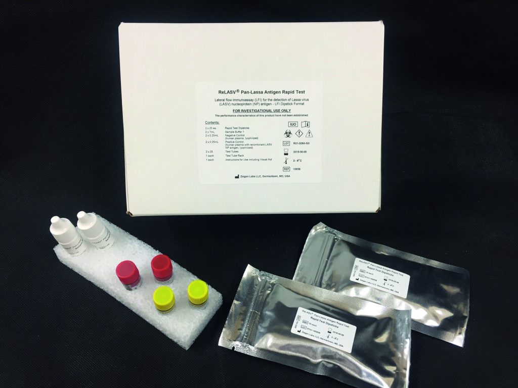
ReLASV® Pan-Lassa Antigen Rapid Test Kit
Antigen and Antibody
Our always vigilant immune system reacts quickly to chase and kill any foreign substance entering our body. These substances – which can be present in bacteria, fungi, or viruses – are known as antigens and are usually the first port of call when scientists are trying to find a target to develop a new vaccine: find a way to enhance this response from the immune system and it will be harder for the invading pathogen to cause disease. The response from the immune system takes the form of antibodies, which are large proteins circulating in the bloodstream always on the look-out for pathogen by recognising and binding to their antigens.
In the Lassa virus, the antigen is a protein complex located in the viral surface. Researchers know this is the protein that the immune system recognises, but the mechanism is still unknown. Keen to unveil the mystery, Viral Hemorrhagic Fever Consortium (VHFC) collaborators at Tulane University and Scripps Research characterised 113 human antibodies derived from Lassa fever survivors and identified 16 that were able to neutralise Lassa virus. It turns out these antibodies can’t spot small sections in the target protein but instead need to recognise its full structure. As this information is vital to design a new vaccine, the researchers had to confirm the results by introducing several mutations which caused minimal conformational changes in the viral protein but severely disrupted antibody activity.
For Dr Boisen, ‘these studies provide the initial foundation for understanding the molecular mechanisms of antibody development, antibody binding and antibody-mediated neutralisation of Lassa in humans. These results will facilitate development of vaccines or antibody-based therapeutics, both urgently needed in this region of the world.’ Dr Boisen is the principal investigator in a new National Institutes of Health grant supporting the development and optimisation of new laboratory tests to measure antibodies resulting from vaccinations. The ReLASV® Pan-Lassa GP Antibody ELISA test will serve as a companion diagnostic in future vaccine studies and aid in Lassa fever surveillance throughout West Africa.
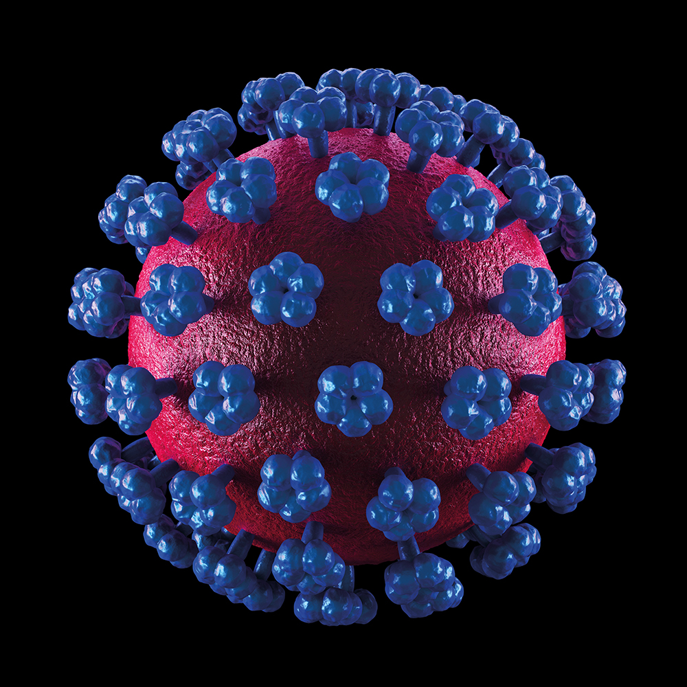
Lassa virus particle
How do Antibodies Neutralise the Lassa Virus?
After determining which antibodies work, the VHFC team wanted to explore how they recognise and neutralise the Lassa virus. ‘Antibodies can be exploited to gain valuable insights into immune responses to pathogens, explains Dr Boisen. ‘Determining the sites at which antibodies bind to pathogens and mediate reaction by the immune system, can reveal mechanisms of immune surveillance, evasion or escape.’
The Scripps Research collaborators quickly realised that the best way to achieve their aim was to study the structure of a virus bound to an antibody. This was not the first time this had been attempted but previous studies had been unsuccessful. The main problem was that, under normal circumstances, using the natural viral protein in vitro always led to unstable changes in conformation. These changes mimic how the protein ‘unfolds’ during the virus invasion of target cells. After 10 years spent trying to solve this problem, Dr Sapphire and her team at Scripps finally identified a few mutations to stabilise the protein to keep its structure prior to binding to target cells (prefusion) without affecting how it binds to the antibody. Interestingly, this approach allowed the identification of certain areas acting as shields, namely on the side and lower portion of the protein, leaving only a few regions vulnerable to antibody binding.
The immune systems antibodies have found a way to maximise their power: this is to act before these shields are put in place, which occurs when the virus enters the target cell. By binding to the viral protein and locking it into the prefusion conformation, the antibody can prohibit viral invasion of the target cells and stop the infection. Indeed, in vitro studies showed a healthy 80% reduction rate in viral activity in the presence of these neutralising antibodies.
‘This structure provides the initial foundation for understanding the molecular mechanisms of neutralisation of the Lassa Virus,’ explains Dr Boisen. Furthermore, it’s possible that other diseases caused by this family of viruses follow a similar pattern of infection and this information may prove valuable to develop new treatments. After all, this antibody also recognises lymphocytic choriomeningitis virus but does not recognise more distantly related Old World Lujo Haemorrhagic fever nor New World arenaviruses.

New Therapeutic Approaches
Several research groups are currently attempting to develop a vaccine, but any breakthroughs may still be years away and Lassa fever patients urgently need a treatment now. From their previous in vitro work, Dr Boisen and his team know that antibodies can target and destroy Lassa viruses. The next obvious step is to test whether the same approach will work in animals.
It turns out that macaques exposed to Lassa virus and then treated with human antibodies didn’t get sick, whereas animals not treated showed a series of symptoms, including problems with liver, spleen, and brain functioning. The researchers further showed that even if treatment was delayed up to a week, when animals were already showing signs of illness, it was still possible to reduce the viral load and clear symptoms.
‘The current studies present a novel alternative for the treatment of Lassa fever infections, using an approach that relies on the use of fully human antibodies derived from convalescent donors,’ adds Dr Boisen. ‘Antibodies targeting the Lassa virus might be of clinical benefit for individuals with Lassa fever, and thus warranted further preclinical study of antibody efficacy.’ Zalgen Labs has developed a human antibody therapeutic (Arevirumab™) which is advancing through preclinical studies in the hope it will eventually be approved as a frontline Lassa fever therapeutic.
Reliable Diagnostic Tools
Another problem that physicians based in remote areas must overcome is that of difficult diagnosis. The laboratory diagnosis of Lassa fever can be achieved using multiple methods but most are not suitable for use as a routine method in the rural endemic regions of West Africa. From Dr Boisen’s perspective, the best possible alternative for use in remote locations relies on detecting the presence of viral antigens in suspected Lassa fever patients using rapid diagnostic tests (RDTs). Similar types of tests are already used to diagnose malaria, dengue, AIDS, and influenza.
Comparing different diagnostic methods, the team showed that Zalgen’s ReLASV® RDT was at least as good as any other standard method, and in some cases, even surpassed the performance of other molecular testing methods such as polymerase chain reaction. The good news is that RDTs do not rely on any equipment and can be completed quickly and with limited resources. ‘Widespread implementation could provide health care workers at remote locations with a reliable diagnostic tool rather than reliance on the less accurate non-specific clinical symptoms that have been the cornerstone of Lassa fever diagnosis for decades,’ notes Dr Boisen.
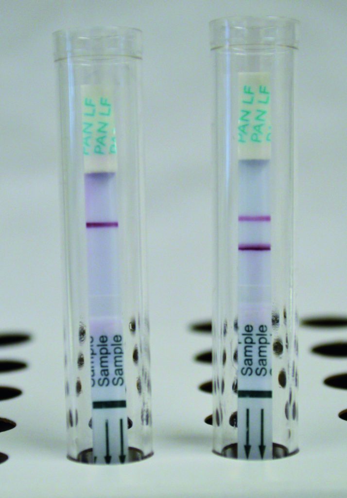
Developed ReLASV RDT assays. Negative result (left) and Positive result (right). Control Line signal (top line) indicates correctly developed rapid test. Test Line signal (bottom line on positive) indicates detection of Lassa virus nucleoprotein antigen.
How to Diagnose and Treat a Patient
The Kenema Government Hospital in Sierra Leone, and the Irrua Specialist Teaching Hospital in Nigeria, are the only two facilities in the world continuously testing for and admitting Lassa fever patients. This is an area well-known to Dr Boisen – the team evaluated the field performance of their ReLASV® RDT with individuals suspected of having Lassa fever from the Kenema Hospital and from control populations living in this area.
Upon arrival at the hospital, if patients present with suspected symptoms, they’re screened for Lassa using Zalgen’s RDT. This provides an initial diagnosis and allows physicians to start ribavirin treatment for positive results. If the result is negative, there is a second approach with more time-consuming methods to confirm the original result.
The benefits of Dr Boisen’s research are thus already apparent with the translation of work to date from lab to clinic bringing practical benefits to the care of Lassa fever patients. Efforts such those undertaken by Dr Boisen and his team hold much-needed promise for reducing the burden and devastating impact of Lassa fever in West Africa.
Reference
https://doi.org/10.33548/SCIENTIA373
Meet the researcher

Dr Matthew Boisen
Director of Diagnostics Development
Zalgen Labs, LLC,
Aurora, CO
USA
After completing a PhD in Biomedical Science from Tulane University in 2015, Dr Boisen joined Zalgen Labs as the Director of Diagnostics Development. The researcher has successfully obtained and managed multiple grants as a principal investigator, including management of international clinical research in collaboration with the Viral Hemorrhagic Fever Consortium (VHFC.org). With over 25 years’ experience, Dr Boisen has unparalleled expertise in the development of rapid, accurate, point-of-care diagnostic tests for infectious diseases and bio-warfare pathogens.
CONTACT
W: http://www.zalgenlabs.com/; www.vhfc.org
KEY COLLABORATORS
Dr Robert F Garry, Tulane University
James E Robison MD, Tulane University
John Schieffelin MD, Tulane University
Donald Grant MD, Kenema Government Hospital, Sierra Leone
Dr. Christian Happi, Redeemers University, Nigeria
Dr Luis M Branco, Zalgen Labs LLC
Dr Erica Olmann-Saphire, La Jolla Institute for Allergy and Immunology
Dr Kathryn M Weinell, La Jolla Institute for Allergy and Immunology
Dr Thomas W Geisbert, Galveston National Lab, University of Texas Medical Branch
Dr Robert W Cross, Galveston National Lab, University of Texas Medical Branch
FUNDING
National Institute of Allergy and Infectious Diseases (NIAID)
Coalition for Epidemic Preparedness Innovation (CEPI)
Bill & Melinda Gates Foundation
FURTHER READING
ML Boisen, et al, Field validation of recombinant antigen immunoassays for diagnosis of Lassa fever, Scientific Reports, 2018, 8, 5939.
KM Hastie, et al, Structural basis for antibody-mediated neutralization of Lassa virus, Science, 2017, 356, 923–928.
C Mire, et al, Human-monoclonal-antibody therapy protects nonhuman primates against advanced Lassa fever, Nature Medicine, 2017, 23, 1146–1149
J Robinson, et al, Most neutralizing human monoclonal antibodies target novel epitopes requiring both Lassa virus glycoprotein subunits, Nature Communications, 2016, 7, 11544.
![]()
Creative Commons Licence
(CC BY 4.0)
This work is licensed under a Creative Commons Attribution 4.0 International License. 
What does this mean?
Share: You can copy and redistribute the material in any medium or format
Adapt: You can change, and build upon the material for any purpose, even commercially.
Credit: You must give appropriate credit, provide a link to the license, and indicate if changes were made.
More articles you may like
International Isocyanate Institute | TDI-induced Asthma: Reanalysing Data to Find Hidden Trends
Even if you’ve never heard of them, you’ve used polyurethanes. Producing them requires toluene diisocyanates, which may/can induce asthma when inhaled. A 5-year study claimed to conclude that cumulative TDI exposure over time was indicative of asthma incidence. However, a reanalysis by a team at the International Isocyanate Institute points the finger instead at the frequency of unprotected high-exposure events, like accidental spills or plant maintenance. This finding guides the way for future advances in worker safety.
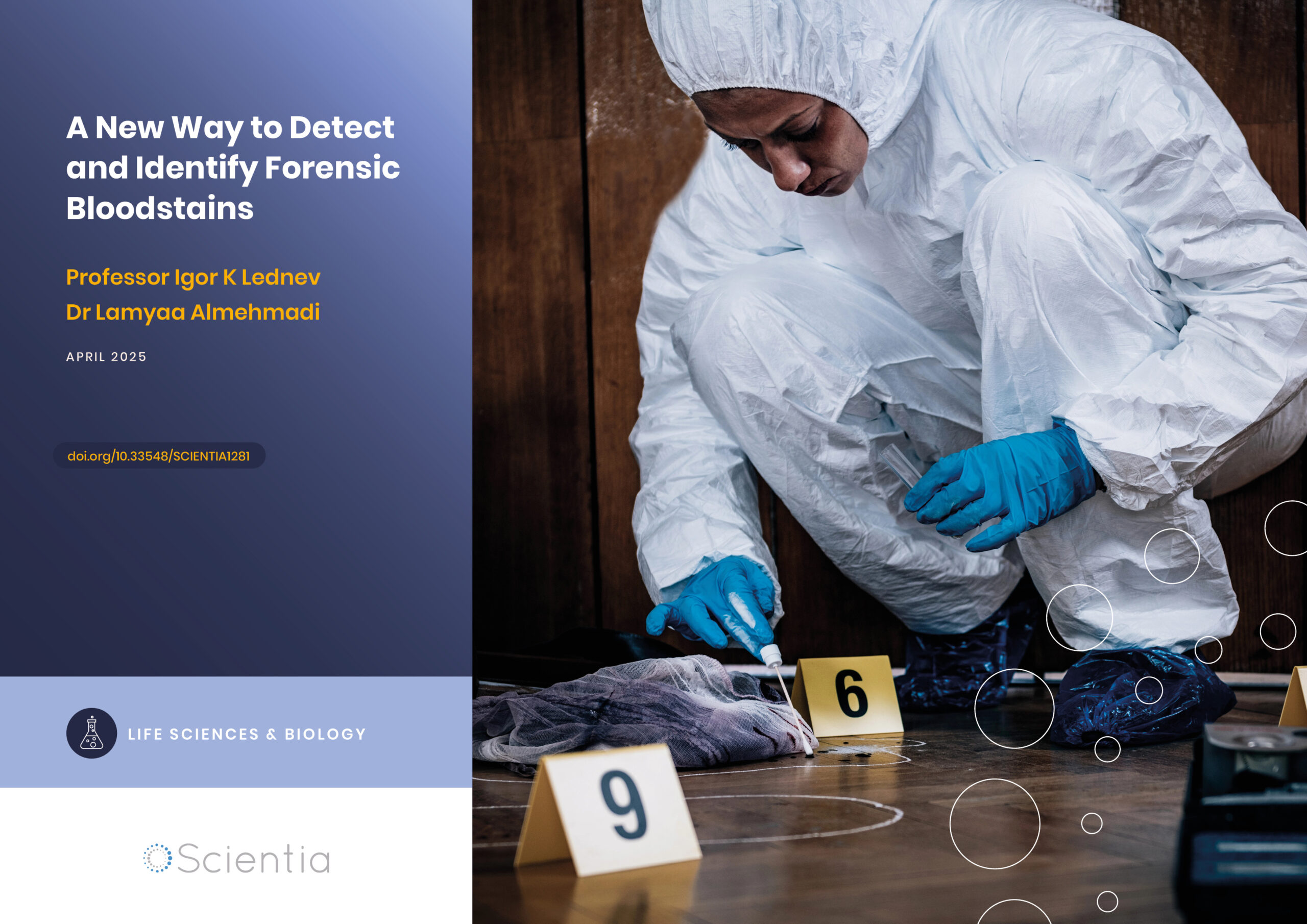
A New Way to Detect and Identify Forensic Bloodstains
Accurately identifying bodily fluids at crime scenes is vital to aid forensic examinations and obtain information for use in criminal proceedings. However, collecting viable material for analysis can be challenging, especially if samples are difficult to access or the amount is minute. Dr Lamyaa Almehmadi and Professor Igor K Lednev at the University at Albany, State University of New York, USA, have introduced a new technique to assist in analysing bloodstains for forensic examination without compromising sample integrity.
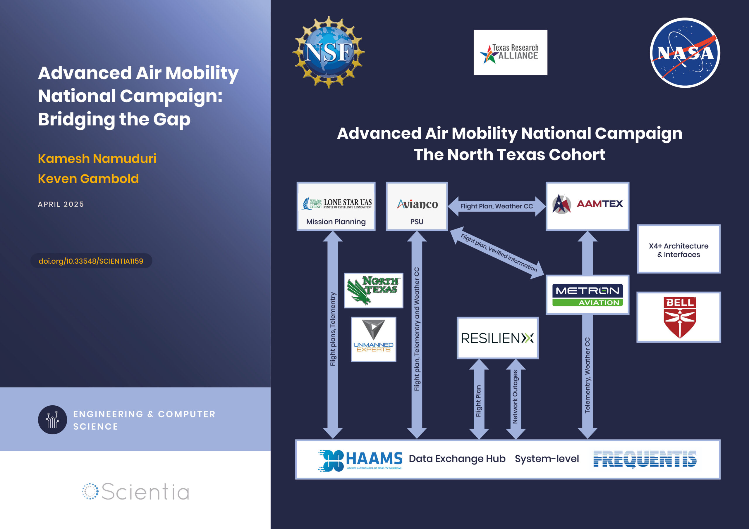
Advanced Air Mobility National Campaign: Bridging the Gap
The rapid advancements in technology have paved the way for revolutionary changes in transportation, particularly air mobility. One such groundbreaking initiative is the Advanced Air Mobility (AAM) National Campaign led by NASA. This campaign aims to integrate advanced air mobility solutions into the existing transportation infrastructure, creating a seamless, efficient, and safe urban air transportation system. By addressing the various challenges associated with urban air mobility, the AAM National Campaign is poised to redefine how we navigate our cities, ultimately leading to reduced congestion, improved accessibility, and enhanced environmental sustainability.
Dr Niloofar Vardian | Mapping the Unknown: Inside Black Holes
Dr Niloofar Vardian at the SISSA school has advanced our understanding of black hole interiors through precise mathematical modelling. Her recent publication sheds light on previously inaccessible aspects of black hole dynamics, deepening our knowledge of these mysterious and difficult-to-study phenomena.