
Dr Muy-Teck Teh – A Novel Diagnostic Tool for Cancer Detection
Head and neck squamous cell carcinoma (HNSCC) constitutes around 90% of all head and neck cancers. Millions of individuals are diagnosed across the globe every year, very often too late and with poor prognosis. Among other factors, alcohol consumption and smoking increase the risk to develop HNSCC. Dr Muy-Teck Teh, from Queen Mary University of London, is driving forward our understanding of the factors leading to cancer, leading the development of novel less invasive detection methods, and progressing better therapeutic options.
Early Detection is Key
The early detection of precancerous lesions remains the most efficient way to prevent cancer development and minimises the risk of intensive surgery. This is important, because surgical treatments may lead to physical disfiguration or functional handicaps, such as impaired swallowing or breathing, both of which can significantly impact on the patient’s overall quality of life.
The current method of identifying a cancerous, malignant lesion is based on the microscopic observation of the tissue. This histopathology is a costly, time-consuming and unreliable procedure for detecting an early tumour. It requires invasive biopsies to obtain a tissue sample 5–20mm in size, large enough so that the pathologist can observe the difference between the malignant and healthy cells. It often requires suturing, causing significant pain to the patient. The accurate observation of the malignant cells is highly dependant on the pathologist’s skills and the preparation of the sample. Furthermore, the diagnostic report can take up to a week to complete, and this waiting may cause extreme stress to the patient. There is an urgent need for rapid and reliable detection methods of early cancer to improve patient outcomes and reduce public healthcare costs.
Dr Teh at Queen Mary University of London is a real-life cancer detective with a career spanning over 20 years. He is committed to understanding the mechanisms underlying the transformation of an abnormal cell growth into cancer, a process known as oncogenesis. In 2019, together with his team, Dr Teh patented the ‘quantitative Malignancy Index Diagnostic System’ (qMIDS), the first diagnostic test for the early detection of oral cancers. Being 90% more accurate than conventional tests and providing results as quickly as in 90 minutes, qMIDS represents a significant step forward in the early detection of cancer. Work by Dr Teh is ongoing to take this even further.

FOXM1: Molecule of the Year
Most cancers occur as a result of DNA alterations such as mutations, amplifications or deletion of genetic material leading to uncontrolled cell proliferation. Over the last decades, scientists have identified the genes and proteins that are expressed during the different phases of tumour development. However, the clinical use of these data remains a scientific challenge. Although biomarkers for certain types of cancer have already been identified, head and neck cancers remain very difficult to diagnose.
Dr Teh and his colleagues concentrate their research on the Forkhead box protein M1 (FOXM1), a human protein coded by the gene FOXM1 and influences cell fate such as division or death, therefore playing an important role in regulating cell’s ability to proliferate. In fact, Dr Teh and his group were the first to provide evidence for the role of FOXM1 in human cancer. Since their seminal work published in 2002, the field of FOXM1 expanded exponentially and it is now a key oncogene that is found to be driving cancer progression in almost all human cancer types.
FOXM1 belongs to a group of proteins called transcription factors, able to bind DNA and regulate the transcription of a DNA sequence into messenger RNA (mRNA). mRNA then exits the cell nucleus to be translated into proteins. FOXM1 is particularly relevant in cancer research as it regulates numerous genes involved in different stages of the disease, from initiation to metastasis. FOXM1 was designated Molecule of the year in 2010 by the International Society for Molecular and Cell Biology and Biotechnology Protocols and Research for its potential in cancer research. Dr Teh and his colleagues use FOXM1 as a ‘molecular gauge’ to quantify the progression of cancer in single tissue biopsy.

A key role for FOXM1 in the regulation of stem cell renewal was unveiled by Dr Teh. Abnormal activity of FOXM1 leads to excess stem cell renewal and subsequently promoting tumour initiation. Credit Dr Teh.
Quantifying Tumour Progression
Cancers are, unfortunately, very complex diseases and one marker alone would not be sufficiently reliable or accurate for diagnosis. Initially testing 200 potential genes, Dr Teh and colleagues identified 14 relevant FOXM1 associated genes that were expressed differently during cancer and two reference genes, expressed at constant levels. Using real-time polymerase chain reaction, a very reliable and easy technique widely used in laboratories to quantify gene expression, they computed the results into an algorithm to generate a ‘qMIDS malignancy index scoring system’. The score, based on the expression of the 16 genes, is correlated with tumour progression. ‘The qMIDS assay objectively measures the malignancy status of a biopsy tissue sample using molecular signatures of multiple FOXM1-orchestrated biomarkers’ explains Dr Teh.
To demonstrate proof of concept, the accuracy of qMIDS was tested in benign and malignant biopsies from two cohorts of patients from the UK and Norway. The high sensitivity of the test prompted Dr Teh and his colleagues to further investigate if qMIDS could be used to further characterise the tumour. They performed macro dissection of larges tumours and precancerous lesions to compute information and create a malignancy ‘heat map’ based on the molecular information. The heterogeneity of the tumour and the clinical significance of the molecular patterns warrant further investigations, but heat maps allow the simultaneous detection of tumour progression and tumour margin (where the tumour stops), which is highly relevant for surgery.
The next step was to validate the diagnostic in a larger non-European cohort. This was essential as the genetic expression can differ between different ethnic groups. Previous studies have reported that sociodemographic factors can influence the genetic background of HNSCC. Dr Teh and his team tested qMIDS in a Chinese cohort and examined the correlation between qMIDS score and progression to cancer. The study, published in 2016, revealed identical datasets between the European and Chinese populations and further demonstrated the robustness of qMIDS in accurately diagnosing HNSCC in different ethnic groups.
Dr Teh’s additional collaborations with India and Pakistan further provided independent evidence that the pathophysiology of OSCC was molecularly indistinguishable between the Asian and European specimens. The qMIDS test robustly quantifies a universal FOXM1-driven oncogenic program in OSCC which transcends ethnicity, age, gender and geographic origins
Dr Teh and his colleagues continue their optimisation of qMIDS and are now investigating whether qMIDS can also diagnose other types of cancer. They also have promising evidence that qMIDS could be used for the detection of vulva and skin cancers.

The novel, affordable, high-throughput, quantitative Malignancy Index Diagnostic System (qMIDS) can accurately differentiate between low and high-risk oral lesions. Credit Dr Teh.
Cancer Biomarkers Hidden in Body Fluids
Recent studies suggest that exosomes contribute to the development of tumours. Exosomes are very small vesicles formed and released by all cell types. Among other important functions, they transport information from a cell to another. The possibility that cancer cells may use exosomes to send reprogramming signals to other cells contributing to the development of tumour and cancer spread, caught the attention of Dr Teh. The presence of exosomes in body fluids such as blood or saliva represents a promising approach to develop non-invasive diagnostics and therapeutics.
Saliva is a complex body fluid, and the challenge was to identify a single protein that can be used as an exosomal biomarker. Based on their previous observation that the CEP55 protein is regulated by FOXM1, Dr Teh and his colleagues demonstrated that CEP55 is exclusively found in the exosomes of malignant cell culture but is absent in healthy cultures. Further in vivo validations of the results in clinical samples are required but these results, published in 2018, provide confidence that CEP55 up-regulation could be used as an exosomal cancer biomarker.

CEP55 protein localisation using immunogold transmission electron microscopy on exosomes derived from normal human plasma, OK113, SVFN8 and SqCC/Y1. Credit Dr Teh.
Personalised Therapeutics and Future Care
Unfortunately, progress in the treatment of HNSCC is held back by the heterogeneity of tumours and the complexity of the structures they affect. Despite the numerous on-going clinical trials and therapeutic advancements, the survival rate for patients with HNSCC remains too low. Unlike other types of cancer such as breast or lung cancers, HNSCC cancers are treated with a standard combination of treatments regardless of the genetic biomarkers. It is therefore essential to classify HNSCC patients and propose a more tailored plan of intervention. This can prevent unnecessary and aggressive treatments for some patients and alleviate the intervention cost.
In 2019, Dr Teh and his colleagues conducted a retrospective analysis linking sociodemographic and clinicopathological data, allowing the identification of two subgroups of HNSCC patients which were molecularly and clinically distinct. The two opposite molecular signatures (+q6 and -q6) match two well-studied high-risk groups in the UK population, statistically differing in age, sex, ethnicity and lifestyle. For example, the group +q6 had a higher alcohol consumption that the -q6 group. Although further investigations are needed to link the data with tumour progression, the identification of the two subgroups represents a significant step towards personalised molecular-signature-guided treatments for HNSCC patients.
It should be noted that although FOXM1 expression is a powerful tool that can be utilised for diagnostic and therapeutic aims, many individual factors remain to be overcome. FOXM1 can be expressed in at least five confirmed variants and a further seven predicted variants have been identified. Although most people study FOXM1B and FOXM1 C in cancer aetiology, other isoforms are worthy of exploration.
Over 20 years, Dr Teh has made tremendous strides forward in the detection and treatment of HNSCC. Looking to the future, he envisages that patient care will involve combinations of non-invasive oral cancer detection (such as using saliva or blood) to screen asymptomatic patients, and then non-invasive optical or imaging approaches to inform as to the best sampling location. This could then be followed by molecular and histopathological analysis methods to determine an accurate diagnosis and to tailor the most appropriate treatment intervention for patients.
Reference
https://doi.org/10.33548/SCIENTIA572
Meet the researcher

Dr Muy-Teck Teh
Senior Lecturer
Barts & the London School of Medicine & Dentistry
Queen Mary University of London
London
UK
Dr Muy-Teck Teh obtained a BSc (Hons) in Biomedical Science in 1996, followed by a PhD in Physiology, from King’s College London, in 2000. He undertook two postdoctoral research positions, funded by the Wellcome Trust and then Cancer Research UK. Dr Teh is now a Senior Lecturer in Head and Neck Cancer at Barts & the London School of Medicine & Dentistry, Queen Mary University of London. As part of his outstanding research career to date, Dr Teh pioneered the identification of FOXM1 as a key driver in human cancer initiation which was awarded ‘Molecule of the Year’ in 2010 by the International Society for Molecular and Cell Biology and Biotechnology Protocols and Research. He leads a research group investigating cancer biomarkers and novel diagnostic methods with the overarching aim of personalising cancer treatment based on individual molecular signatures. In 2019, he patented the world first FOXM1-based digital molecular cancer test ‘quantitative malignancy diagnostic system (qMIDS)’ for the early detection of oral cancer. Dr Teh has numerous international collaborators across the world and has published over 60 papers in prestigious journals.
CONTACT
E: m.t.teh@qmul.ac.uk
W: http://www.dentistry.qmul.ac.uk/people/profiles/drmuyteckteh.html
Twitter: https://twitter.com/FOXM1B
ORCID: https://orcid.org/0000-0002-7725-8355
Laboratory: https://sites.google.com/view/qmids/home
KEY COLLABORATORS
Edward W Odell, King’s College London, UK
Allan Hackshaw, University College London, UK
Eric Lam, Imperial College London, UK
Christain Simon, University of Lausanne, Switzerland
Daniela E. Costea, University of Bergen, Norway
Dipak Sapkota, University of Oslo, Norway
Bengt Hasséus, University of Gothenburg, Sweden
Monica C Solomon, Manipal University, India
Akhilanand Chaurasia, King George’s Medical University, India
Malik Waqar Ahmed, Comsats University, Pakistan
Hong Ma, Guizhou Medical University, China
Hao Chen, Guangzhou Medical University, China
William A. Yeudall, Augusta University, USA
M. Tilakratne, University Malaya
FUNDING
Medical Research Council
Wellcome Trust
Saving Faces
The Rosetrees Trust
State Administration of Foreign Experts Affairs, China
Guizhou Medical University
Union for International Cancer Control
Pakistan Higher Education Commission
Guangzhou Medical University, Cancer Hospital and Institute
British Council
FURTHER READING
F Qadir, A Lalli, HH Dar, et al, Clinical correlation of opposing molecular signatures in head and neck squamous cell carcinoma, BMC Cancer, 2019, 19, 830.
F Qadir, MA Aziz, CP Sari, et al, Transcriptome reprogramming by cancer exosomes: identification of novel molecular targets in matrix and immune modulation, Molecular Cancer, 2018, 17, 97.
MT Teh, Oral Cancer Biomarkers: Is it a Meaningless Game? The Open Dentistry Journal, 2016, 3, e1–e3.
H Ma, H Dai, X Duan, Z Tang, et al, Independent evaluation of a FOXM1-based quantitative malignancy diagnostic system (qMIDS) on head and neck squamous cell carcinomas, Oncotarget, 2016, 7, 54555–54563.
MT Teh, Can Challenges of Oral Cancer Diagnosis be Resolved? Dental Health Current Research, 2015, 1.
Teh MT, Is salivary exosome the answer to early detection of oral cancer? The Open Dentistry Journal, 2015, 2, e3–e4.
MT Teh, IL Hutchison, DE Costea, et al, Exploiting FOXM1-orchestrated molecular network for early squamous cell carcinoma diagnosis and prognosis, International Journal of Cancer, 2013, 132, 2095–2106.

Want to republish our articles?
We encourage all formats of sharing and republishing of our articles. Whether you want to host on your website, publication or blog, we welcome this. Find out more
Creative Commons Licence
(CC BY 4.0)
This work is licensed under a Creative Commons Attribution 4.0 International License. 
What does this mean?
Share: You can copy and redistribute the material in any medium or format
Adapt: You can change, and build upon the material for any purpose, even commercially.
Credit: You must give appropriate credit, provide a link to the license, and indicate if changes were made.
More articles you may like

Dr Lifei Wang | Can Species Distribution Models Inform Us About Future Ecosystems?
The world is buzzing with news about how human activities and climate shifts are reshaping our ecosystems. Have you ever wondered how life will adapt to this rapidly changing world? Ecologists might be able to predict how different species will live in future using computer simulations. Dr Lifei Wang at the University of Toronto Scarborough investigates how different stimulations work under varying conditions to provide new insights into what may lie ahead.
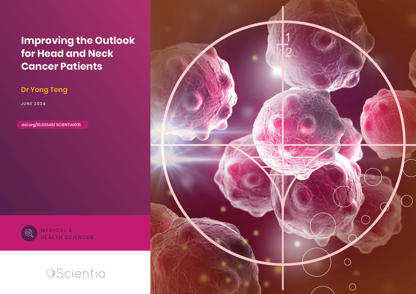
Dr Yong Teng | Improving the Outlook for Head and Neck Cancer Patients
Dr Yong Teng at the Emory University School of Medicine is working with colleagues to overcome the high mortality of individuals diagnosed with cancers affecting the head and neck. One of his approaches is based on understanding the particular mechanisms of the ATAD3A gene, which new insights suggest are closely related to cancers affecting the head and neck.
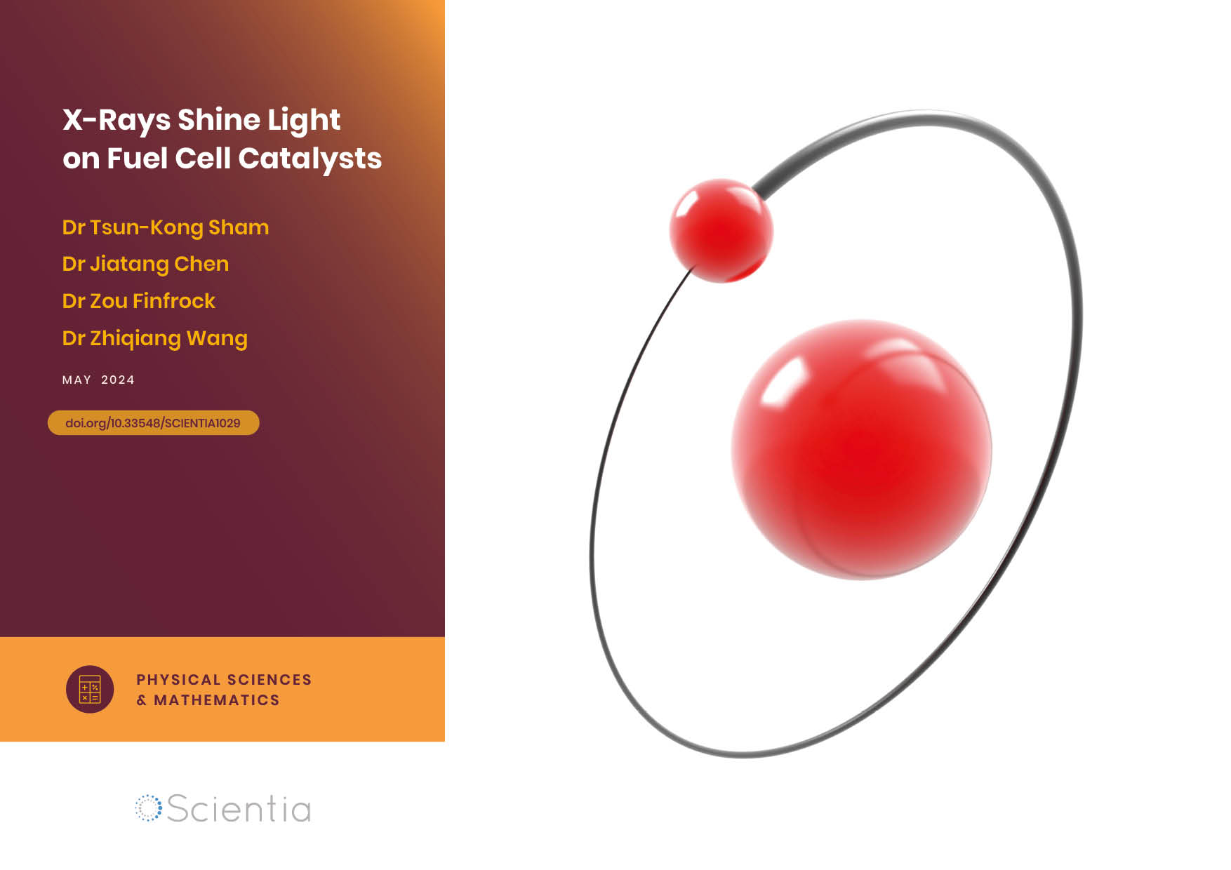
Dr Tsun-Kong Sham – Dr Jiatang Chen – Dr Zou Finfrock – Dr Zhiqiang Wang | X-Rays Shine Light on Fuel Cell Catalysts
Understanding the electronic behaviour of fuel cell catalysts can be difficult using standard experimental techniques, although this knowledge is critical to their fine-tuning and optimisation. Dr Jiatang Chen at the University of Western Ontario works with colleagues to use the cutting-edge valence-to-core X-ray emission spectroscopy method to determine the precise electronic effects of altering the amounts of platinum and nickel in platinum-nickel catalysts used in fuel cells. Their research demonstrates the potential application of this technique to analysing battery materials, catalysts, and even cancer drug molecules.
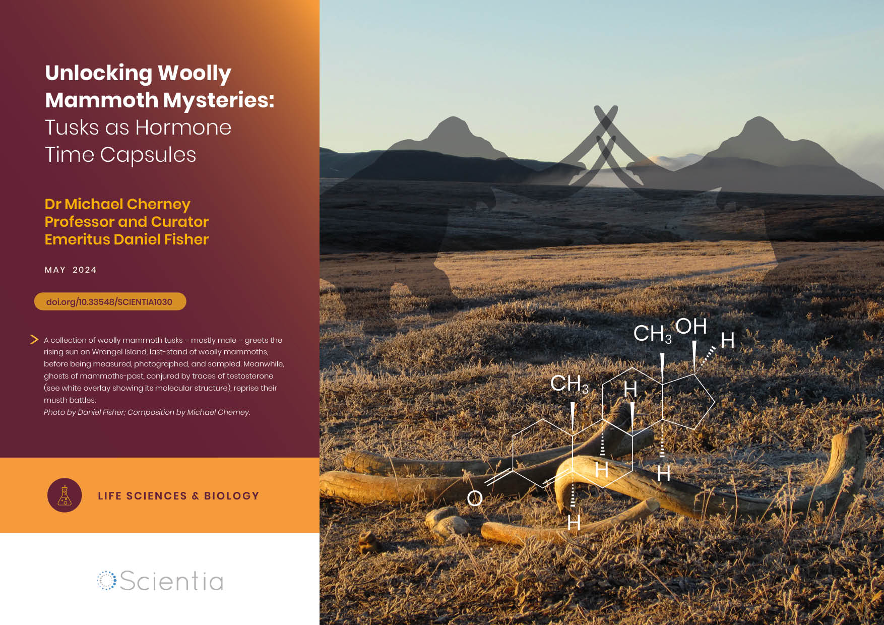
Dr Michael Cherney – Professor Daniel Fisher | Unlocking Woolly Mammoth Mysteries: Tusks as Hormone Time Capsules
The impressive tusks found on proboscideans (the order of mammals that includes elephants, woolly mammoths, and mastodons) are like time capsules, preserving detailed records of their bearers’ lives in the form of growth layers and chemical traces. Frozen in time for thousands of years, these layers can unlock secrets about the lives of long-extinct relatives of modern elephants. Dr Michael Cherney and Professor Daniel Fisher from the University of Michigan used innovative techniques to extract and analyse steroid hormones preserved in woolly mammoth tusks. This ground-breaking work opens new avenues for exploring the biology and behaviour of extinct species.