
Dr Robyn S. Klein – Regulation and Loss of Neuroprotection in Viral and Autoimmune Diseases of the Central Nervous System
Viral and autoimmune diseases of the central nervous system (CNS) are often characterised by the onset of inflammation leading to neurological dysfunction, including impairment to memory and other cognitive domains. Dr Robyn S. Klein at the Washington University School of Medicine in St. Louis, leads a team that specialises in neuroinflammatory diseases of the CNS. In recent years, they have investigated the regulation of blood-brain barrier permeability in autoimmune diseases and viral infections with pathogens such as the West Nile virus.
Blood-brain Barrier Permeability in Autoimmune and Viral Diseases
Dr Robyn S. Klein is an internationally recognised expert in the field of inflammatory diseases of the central nervous system (CNS). In recent years, her laboratory has been focusing on the molecular mechanisms behind inflammation and how they regulate blood-brain barrier (BBB) permeability in viral and autoimmune diseases.
Dr Klein’s team has developed in vitro (outside the living organism) models of the BBB to investigate what mechanisms allow pathogens like the West Nile Virus (WNV) and white blood cells released during autoimmune disease to gain access to the CNS, causing encephalitis. The team has also used in vivo (within the living animal) models of autoimmune and WNV encephalitis to identify the molecular basis of persistent inflammation.
The BBB is composed of microvascular endothelial cells joined by tight junctions, the latter consisting of regulatory proteins that control cellular permeability. Viruses may cross the BBB through several routes, including direct infection of the microvascular endothelial cells of the brain and infiltration of infected peripheral white blood cells. Viral trafficking across the BBB is also possible as a result of increased permeability of the endothelium, which in turn can be a consequence of viral attack mechanisms or the overexpression of inflammatory cytokines, such as tumour necrosis factor alpha (TNF-α) and interleukin-1 (IL-1).

The Link Between Cytokine Signalling and Neuroinflammation
In a study published in 2014, Dr Klein and her collaborators highlighted how WNV, a mosquito-borne pathogen that causes viral encephalitis, triggers the expression of TNF-α and IL-1 to cause a cascade of reactions that limit viral replication, facilitate antigen presentation for recognition by the immune cells, and direct the trafficking of antiviral white blood cells. In the same study, they showed that despite limiting viral replication in the CNS, TNF-α and IL-1 signalling can also contribute to neuropathology by directly enhancing mice BBB permeability in vivo (that is, within the living organism) and indirectly facilitating the trafficking of WNV across the endothelium.
In contrast with the BBB disruption caused by the above-mentioned cytokines, the 2014 study showed that a different cytokine, known as type I interferon (IFN), promoted and stabilised BBB function instead. This finding was supported by experiments in vivo, since mice with impaired type I IFN responses exhibited enhanced BBB permeability. In the same study, the researchers demonstrated that promoting Type-I IFN signalling in vitro resulted in an enhancement of the barrier function over baseline conditions rescuing Il-1 and TNF- α-mediated barrier dysregulation, suggesting that CNS Type-I IFN may be able to reverse BBB permeability. These findings were followed by the discoveries that type III interferon and the TAM receptor Merk can each synergise with type I interferon in maintaining BBB integrity during neurotropic viral infections.
Synaptic Loss During NWV Infection: The Role of Astrocytes
Although inflammation is necessary for CNS infection clearance, immune molecules can trigger a cascade of events that lead to neurological dysfunction. Patients surviving WNV encephalitis show high rates of memory impairment and cognitive impairment. More specifically, detriments to neurons in the hippocampus, which is essential for visual and spatial (visuospatial) processing – the ability to identify visual and spatial relationships among objects – and visuospatial memory, are commonly found.
In 2016, Dr Klein and her colleagues published a study demonstrating their development of an effective model of WNV-induced spatial memory impairment. Critically, this allowed them to identify that the virus induces elimination of presynaptic terminals without loss of hippocampal neurons or volume, pointing to this as a potential mechanism underlying such deficits in patients recovering from WNV infection.
The Klein laboratory later published a study in 2017 confirming that IL-1 alters the proliferation and differentiation of neural progenitor cells, which cause an increase in the production of astrocytes and a decrease in the production of neurons. Astrocytes are star-shaped cells that surround and outnumber neurons in the CNS. They have an important role in the maintenance of the BBB, bridging the neural tissue to the vascular tissue and secreting factors that regulate the inflammatory cascade during infection.
It is plausible that pro-inflammatory astrocytes may contribute to the start of a vicious circle following infection with NWV, by triggering the recruitment of IL-1, which in turn continues to inhibit neurogenesis, causing the memory impairment and loss of hippocampal synapses observed during WNV neuroinvasive disease. This was confirmed by in vivo results showing that, following infection with WNV, genetically modified mice lacking the ability to express IL-1 exhibit a significant recovery in the number of synapses and in their cognitive abilities in comparison with wild type animals as found in nature.

Zika virus in blood
Interferon-γ Regulates Neuron Loss During WNV and Zika Virus Infection
The recently emerged flavivirus Zika Virus (ZIKV) can cause several neurological complications in adults, including encephalopathy, meningomyelitis and encephalomyelitis, which can affect memory and cognition with unknown long-term outcomes. Recovery from infection with flaviviruses is associated with reduced learning abilities, suggesting that there might be an underlying mechanism of cognitive impairment that correlates with a broader category of memory disorders.
Activation of microglia and astrocytes in the brain during acute infection promotes the recruitment of antiviral T cells, via a cytokine-mediated mechanism. T-cell cytokines such as IFN-γ (not to be confused with the Type-I IFN discussed above) may also influence microglial biology, as observed in brain tissues of patients with multiple sclerosis (MS), Parkinson’s disease and Alzheimer’s disease.
Dr Klein and her team reported in a paper published in 2019 that antiviral T cells persist within the hippocampus after recovery from flaviviral infection in two in vivo animal models of WNV and ZIKV. The presence of T cells is associated with the development of specific learning defects through microglial activation and loss of synapses formation. The two pathogens, however, caused slightly different outcomes; while WNV was responsible for the elimination of presynaptic termini, ZIKV promoted the loss of neuronal nuclei and postsynaptic termini. As further confirmation of this, animals deficient in CD8+ T cells or IFN-γ signalling in microglia demonstrated protection against synapse elimination following WNV infection and decreased neuronal apoptosis with synapse recovery following ZIKV infection.

Type I-Interferon Protects the BBB in Viral Equine Encephalitis
Venezuelan and western equine encephalitis viruses (VEEV and WEEV, respectively) and eastern equine encephalitis virus (EEEV) are emerging infectious diseases in the Americas, causing several major outbreaks in the human and horse population during the past few decades. Shortly after infection, these viruses can infect the CNS, resulting in severe long-term neurological deficits or death.
Following peripheral infection with alphaviruses, VEEV and WEEV are trafficked at the BBB into the CNS. The transmigration occurs via a caveolin-1 (Cav-1)-mediated mechanism across an intact BBB, which can be halted by Type I-IFN.
In vivo examination of early viral entry in Cav-1-deficient mice confirmed the presence of significantly lower VEEV and WEEV viral burdens in the brain in comparison to similarly infected wild-type animals, confirming that Cav-1 signalling allows alphaviruses to enter the CNS and that type-I IFN limits this process at the BBB.

Three types of brain cells. Red: astrocytes, green: pyramidal
neurons, blue: microglia cells
Mechanisms of Neuroinflammation in Multiple Sclerosis
The autoimmune disease MS and one of its animal models, experimental autoimmune encephalomyelitis (EAE) are characterised by a pronounced T cell autoimmune response against the myelin sheath that protects the nerves in the CNS, leading to motor and sensory function loss. Dr Klein and her team, following previous reports that IL-1, TNFα and other cytokines have critical roles in MS and EAE (including the upregulation of vascular adhesion molecules at the BBB), decided to delve deeper into understanding the molecular mechanisms that facilitate immune cell entry into the CNS during neuroinflammation.
The team investigated cytokine signalling in different regions of the CNS, following induction of EAE in mice, and reported the findings of the study in a paper published in 2020. The study identified a ‘regionality’ in the way cytokines direct T cells towards different parts of the CNS.
Using in vivo models of spinal cord versus brain stem trafficking of myelin-specific T cells, the researchers confirmed that different cytokines differentially regulate astrocyte expression of the adhesion molecules VCAM-1 and CXCR7 in these locations. While the IN-17 cytokine preferentially upregulated VCAM-1 on brain stem astrocytes, IFNγ, modulated the expression of CXCR7 chemokine, facilitating T cell entry into the spinal cord. In the absence of IFNγ, brain stem astrocytes upregulated VCAM-1 expression and astrocyte-specific deletion of VCAM-1 reduced the severity of EAE, suggesting IFNγ may partially limit T cell entry into the brain stem during EAE by inhibiting the expression of VCAM-1 on astrocytes.
Next Steps
It is clear that astrocytes play important roles in neurological diseases at multiple stages of pathogenesis and repair. Astrocytes also critically impact recovery in several models of neurodegeneration, and fragmented mitochondria released by damaged neurons may be removed by neighbouring astrocytes.
There is emerging evidence to suggest that altered astrocyte function may contribute to Parkinson’s disease, but the underlying mechanisms have not been fully elucidated. The Klein laboratory is continuing to investigate the roles played by cytokine signalling and astrocytes in CNS autoimmunity, to provide new insights for the development of CNS-targeted therapies.
Reference
https://doi.org/10.33548/SCIENTIA714
Meet the researcher

Robyn S. Klein, MD, PhD
The Robert E. and Louise F. Dunn Professor of Medical Sciences
Director, Center for Neuroimmunology & Neuroinfectious Diseases
Professor, Departments of Medicine, Pathology & Immunology, Neurosciences
Washington University School of Medicine
St. Louis, MO
USA
Dr Robyn Sue Klein obtained her MD and PhD in Neuroscience in 1993 from the Albert Einstein College of Medicine, New York. After completing clinical training in Internal Medicine, Infectious Diseases and a postdoctoral fellowship in Immunology at Harvard Medical School, she joined Washington University School of Medicine, St. Louis, where she is currently the Robert E. and Louise F. Dunn Professor of Medical Sciences, Director of the Center for Neuroimmunology and Neuroinfectious Diseases, and Professor of Medicine, Pathology & Immunology, and Neurosciences. She has received numerous awards, the most recent being the Distinguished Educator Award, Washington University School of Medicine, St. Louis, and Chair of the NIH Study Section, Clinical Neuroimmunology & Brain Tumors. Dr Klein, who has co-authored more than 100 peer-reviewed publications, was in 2021 appointed as Co-Editor-in-Chief for the prestigious Journal of Neuroimmunology.
CONTACT
E: rklein@wustl.edu
W: https://infectiousdiseases.wustl.edu/faculty-staff/robyn-s-klein/
Twitter: KleinLab_WUSTL
KEY COLLABORATORS
Anne Cross, MD, Washington University School of Medicine
Laura Piccio, MD, PhD, Washington University School of Medicine
John Russell, PhD, Washington University School of Medicine
Gregory Wu, MD, PhD, Washington University School of Medicine
Victor Song, PhD, Washington University School of Medicine
Michael S. Diamond, MD, PhD Washington University School of Medicine
Jonathon Miner, MD, PhD Washington University School of Medicine
Joseph Culver, PhD, Washington University School of Medicine
Mehul Suthar, PhD, Emory University College of Medicine
FUNDING
NIH/NINDS, NIH/NIAID, NMSS, Dana Foundation, DOD
FURTHER READING
M Boldrini, P Canoll, RS Klein, How COVID-19 Affects the Brain, JAMA Psychiatry, 2021, 78(6), 682–683.
L Piccio, GF Wu, RS Klein, A new era in neuroimmunology, Journal of Neuroimmunology, 2021, 351, 577478.
M Kanmogne, RS Klein, Neuroprotective versus Neuroinflammatory Roles of Complement: From Development to Disease, Trends in Neuroscience, 2021, 44(2), 97–109.
RS Klein, Clinical Implications of Basic Research: On complement, microglia, and memory, New England Journal of Medicine, 2020, 382(21), 2056–2058.
H Salimi, MD Cain, X Jiang, et al., Encephalitic Alphaviruses Exploit Caveola-Mediated Transcytosis at the Blood-Brain Barrier for Central Nervous System Entry, mBio, 2020, 11(1), e02731-19.
C Garber, A Soung, LL Vollmer, et al., T cells promote microglia-mediated synaptic elimination and cognitive dysfunction during recovery from neuropathogenic flaviviruses, Nature Neuroscience, 2019, 22(8), 1276–1288.
C Garber, MJ Vasek, LL Vollmer, et al., Astrocytes decrease adult neurogenesis during virus-induced memory dysfunction via IL-1, Nature Immunology, 2018, 19(2), 151–161.
RS Klein, C Garber, N Howard, Infectious immunity in the central nervous system and brain function, Nature Immunology, 2017, 18(2), 132–141.
RS Klein, CA Hunter, Protective and Pathological Immunity During Central Nervous System Infections, Immunity, 2017, 46(6), 891–909.
RS Klein, R Voskuhl, BM Segal, et al., Speaking out about gender imbalance in invited speakers improves diversity, Nature immunology, 2017, 18(5), 475–478.
MV Vasek, C Garber, D Dorsey, et al., A complement-microglial axis is required for synaptic loss during virus-induced memory impairment, Nature, 2016, 534(7608), 538–43.
Lazear, H.M., Daniels, B.P., Pinto, A.K., Huang, A.C., Vick, S.C., Doyle, S.E., Gale Jr., M., Klein, R.S.*, and Diamond, M.S.* (2015) Interferon lambda restricts West Nile virus neuroinvasion by tightening the blood-brain barrier. Sci Transl Med. 22;7(284):284ra59.
JJ Miner, BP Daniels, B Shrestha, et al., The TAM receptor Mertk protects against neuroinvasive viral infection by maintaining blood-brain barrier integrity, Nature Medicine, 2015, 21, 1464–1472.
BP Daniels, DW Holman, LC Cruz-Orengo, et al., Viral pathogen-associated molecular patterns regulate blood-brain barrier integrity via competing innate cytokine signals, mBio, 2014, 5(5), e01476–14.

Want to republish our articles?
We encourage all formats of sharing and republishing of our articles. Whether you want to host on your website, publication or blog, we welcome this. Find out more
Creative Commons Licence
(CC BY 4.0)
This work is licensed under a Creative Commons Attribution 4.0 International License. 
What does this mean?
Share: You can copy and redistribute the material in any medium or format
Adapt: You can change, and build upon the material for any purpose, even commercially.
Credit: You must give appropriate credit, provide a link to the license, and indicate if changes were made.
More articles you may like

Dr Lifei Wang | Can Species Distribution Models Inform Us About Future Ecosystems?
The world is buzzing with news about how human activities and climate shifts are reshaping our ecosystems. Have you ever wondered how life will adapt to this rapidly changing world? Ecologists might be able to predict how different species will live in future using computer simulations. Dr Lifei Wang at the University of Toronto Scarborough investigates how different stimulations work under varying conditions to provide new insights into what may lie ahead.
Dr Yong Teng | Improving the Outlook for Head and Neck Cancer Patients
Dr Yong Teng at the Emory University School of Medicine is working with colleagues to overcome the high mortality of individuals diagnosed with cancers affecting the head and neck. One of his approaches is based on understanding the particular mechanisms of the ATAD3A gene, which new insights suggest are closely related to cancers affecting the head and neck.

Dr Tsun-Kong Sham – Dr Jiatang Chen – Dr Zou Finfrock – Dr Zhiqiang Wang | X-Rays Shine Light on Fuel Cell Catalysts
Understanding the electronic behaviour of fuel cell catalysts can be difficult using standard experimental techniques, although this knowledge is critical to their fine-tuning and optimisation. Dr Jiatang Chen at the University of Western Ontario works with colleagues to use the cutting-edge valence-to-core X-ray emission spectroscopy method to determine the precise electronic effects of altering the amounts of platinum and nickel in platinum-nickel catalysts used in fuel cells. Their research demonstrates the potential application of this technique to analysing battery materials, catalysts, and even cancer drug molecules.
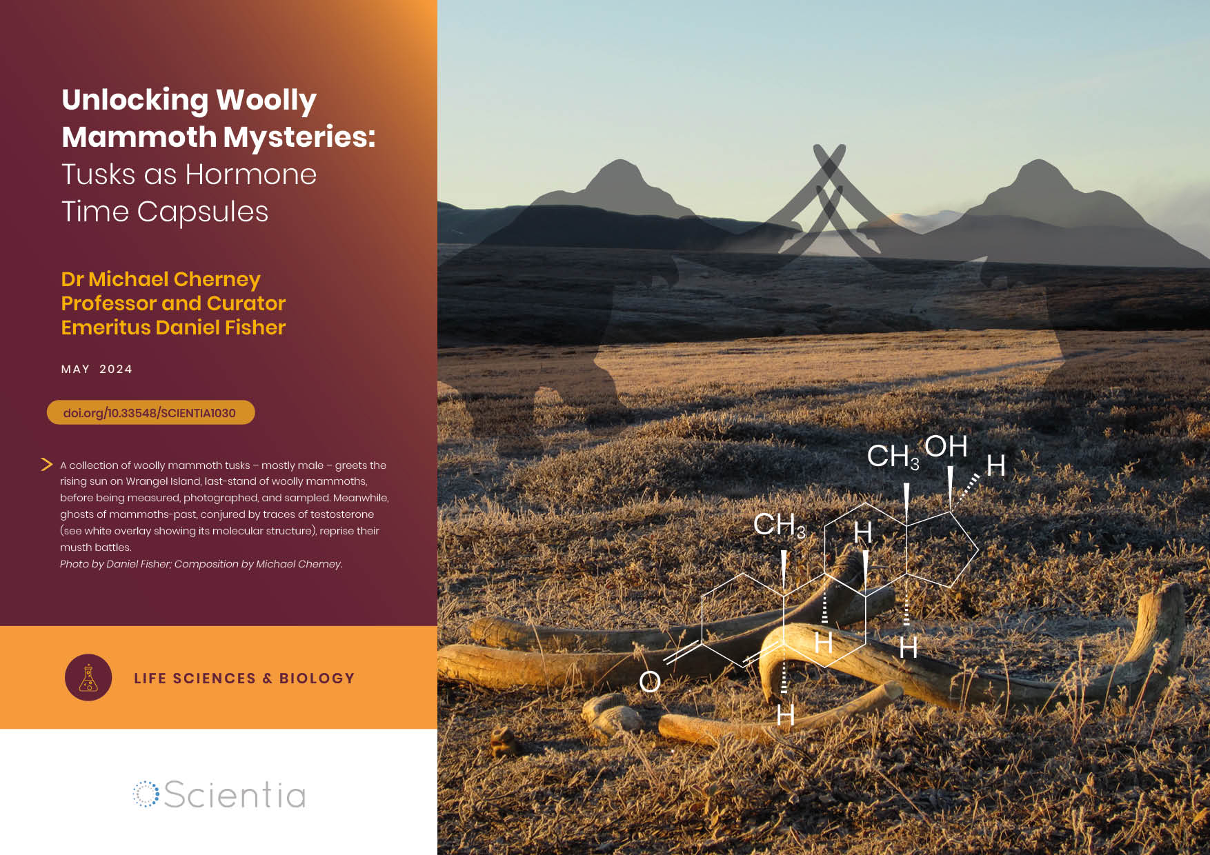
Dr Michael Cherney – Professor Daniel Fisher | Unlocking Woolly Mammoth Mysteries: Tusks as Hormone Time Capsules
The impressive tusks found on proboscideans (the order of mammals that includes elephants, woolly mammoths, and mastodons) are like time capsules, preserving detailed records of their bearers’ lives in the form of growth layers and chemical traces. Frozen in time for thousands of years, these layers can unlock secrets about the lives of long-extinct relatives of modern elephants. Dr Michael Cherney and Professor Daniel Fisher from the University of Michigan used innovative techniques to extract and analyse steroid hormones preserved in woolly mammoth tusks. This ground-breaking work opens new avenues for exploring the biology and behaviour of extinct species.