
Dr Zi-Bing Jin – Building Preclinical Models of Retinal Degeneration in Non-human Primates
Mutations affecting the expression of cone photoreceptors can lead to retinal degeneration, which in many cases can result in a permanent loss of vision. However, preclinical models for human retinal degenerative diseases are lacking. Dr Zi-Bing Jin and his colleagues study rhesus macaque models of achromatopsia (a congenital disorder characterised by an inability to distinguish colours) and oculocutaneous albinism (characterised by a disorder of melanin synthesis, leading to loss of visual acuity). The animal models utilised in the Jin laboratory offer important opportunities for studies on disease mechanisms as well as therapeutic development.
The Macula and the Fovea: High Definition at the Heart of the Eye
Most people are able to see clear, full colour and high-resolution images thanks to the macula, which is located at the centre of the retina. What makes the macula so efficient at detecting light of different wavelengths is the high density of cone photoreceptors expressed in it. Within the central region of the macula lies a ‘pit’ known as the fovea, which is characterised by a higher density of cone photoreceptors and the presence of approximately 25% retinal ganglion cells. A healthy developed fovea plays a critical role in accurate visual acuity.
Mutations affecting the expression of cone photoreceptors can lead to macular degeneration, which in many cases can result in a permanent loss of vision. Achromatopsia is a congenital disorder characterised by an inability to distinguish colours, low visual acuity, excessive sensitivity to light and uncontrolled eye movements. Several gene mutations have been discovered in patients with achromatopsia. The development of the fovea can be seriously hindered by oculocutaneous albinism (OCA), which is characterised by a disorder of melanin synthesis.
Non-human primates have a macula and fovea closely related to those of humans, so scientists can introduce genetic mutations in these mammals to see whether the deletion of specific genes can lead to the loss of cone photoreceptor function. Dr Zi-Bing Jin and his colleagues focus on stem cell translational medicine in retinal health and the genetic mechanisms of eye diseases, and in particular, those affecting the macular and foveal development.
Dr Jin and his team have created a non-human primate model of achromatopsia by partially knocking out a retinal gene known as CNGB3. The gene in question, expressed in the macular cone receptors, is mutated in patients with achromatopsia. The team at the Jin laboratory hopes that the partial knockout of CNGB3 in their animal model could replicate the conditions observed in human patients with achromatopsia. Such a model could then be used in drug development studies and to confirm the involvement of the gene in the development of macular degeneration. The team has also developed rhesus macaque models of OCA to study foveal development and to enable preclinical trials of new therapies for OCA.

A Gene Editing Tool to Investigate the Causes of Achromatopsia
Dr Jin and his collaborators published a paper in 2020 describing the design, development and characterisation of their animal model. They used the CRISPR-Cas9 system, a revolutionary tool for generating mutations, for the macular localised knockout (inactivation) of the CNGB3 gene. The somatic knockout model was initiated by injecting four macaques in the retinal tissue with an adenovirus and the CRISPR-Cas9 system. Following the sub-retinal injection, the team monitored the recovery of the retina at the puncture sites for 30 days. To determine transcriptional changes of CNGB3 in cone photoreceptors, single cells were isolated from the dissected retina for single-cell RNA sequencing (scRNA-seq) studies. A total of 24 cones were picked from the retina and divided into 13 groups, each group containing at least one and a maximum of three cones.
The researchers found that a proportion of treated cells had decreased expressed levels of CNGB3, with a targeting efficiency in infected cells of 12.2%, suggesting that subretinal delivery of adenovirus-mediated CRISPR-Cas9 successfully generated a partial knockout of the gene. After measuring the electrical activity in the retina, the team found that, following the CNGB3 knockout, there was a marked reduction in the expected response from the central retina, suggesting cone dysfunction of the central macula of the primates, consistent with achromatopsia in human patients.
In addition to providing the first non-human primate model for the study of achromatopsia, the paper provided important evidence confirming the safety of CRISPR-Cas9-based gene-editing therapy. The adenoviral-vector-infected areas were only restricted to the retina, leaving other tissues unaltered. This is vitally important in encouraging scientists to perform precise gene editing in vivo. Dr Jin and his collaborators suggested that by improving the targeting efficiency, the CRISPR-Cas9 system could, in future, be used as a ‘gene-editing scalpel’ in the treatment of macular degeneration and other diseases.

Gene Mutations Impairing Melanin Synthesis
The retinal fovea is a region with a very high density of cone photoreceptors, which is responsible for optimal visual acuity in humans. The human fovea contains approximately 25% retinal ganglion cells, and the synaptic connections between cones, bipolar cells, and ganglion cells in a 1:1:1 ratio, resulting in a high level of precision in the transmission of the visual signals.
In primates, melanin synthesis plays an important role in regulating the proliferation and differentiation of retinal cells. Patients presenting with mutations in a gene known as TYR result in the complete loss or partially reduced activity of the amino acid tyrosine, whose chemical modification is the first step in the production of melanin. Mutations in the OCA2 gene also cause a partial impairment in melanin production.
The mechanisms of OCA disease have been reported in many mammal species. However, only non-human primates have a foveal structure similar to that of the human retina. The eye structure of rhesus macaques is very similar to that of the human eye, especially because of the presence of a macula and fovea.
In another paper, published in 2020, Dr Jin and his collaborators reported the development of a rhesus macaque model with spontaneous oculocutaneous albinism, with clinical manifestations similar to those of human OCA patients. Albino macaques presenting with low levels of retinal pigmentation had a measured foveal depth that was significantly shallower than that of healthy subjects. Thicker inner retinal layers at the fovea were also found in the albino subjects.
These observations in rhesus macaques are consistent with oculocutaneous albinism in human patients. Whole-genome sequencing from six macaques showed that all the subjects analysed carried mutations in the OCA2 gene. Additionally, three albino subjects carried another mutation in the TYR gene. The researchers confirmed, via in vitro assays, that both mutations affected the production of melanin.

Driving Further Developments
The model of OCA developed in the Jin laboratory offers new opportunities for studies on disease mechanisms as well as therapeutic development. Dr Jin and his collaborators pointed out in their paper that, although hundreds of mutations in TYR and OCA2 have been reported in OCA patients, only a few studies have included biological assays to validate the effect of the mutations on melanin synthesis. Studies in humans affected by OCA have shown that photoreceptor layers in the fovea continue to grow, albeit at a reduced rate. These reports suggest that earlier-stage treatment might result in a better outcome for these patients. Dr Jin and his team propose that this hypothesis could now be tested in albino rhesus macaques.
In other developments, the Jin laboratory aims to elucidate the disease mechanisms of children with ocular disorders, translating laboratory technology to improve bedside outcomes. Among other ambitious projects under development, Dr Jin and his team are exploring new, groundbreaking ways of growing key ocular tissues from fibroblasts through small molecules and culturing retinal organoids in vitro for the disease modelling of retinitis pigmentosa and retinoblastoma. Following their already highly promising results with the CRISPR-Cas9 system, the team will continue to investigate gene editing for the treatment of complex ocular diseases affecting children and early-onset blindness.
Reference
https://doi.org/10.33548/SCIENTIA698
Meet the researcher

Dr Zi-Bing Jin MD, PhD
Beijing Institute of Ophthalmology
Beijing Tongren Hospital, Capital Medical University
Beijing
China
Dr Zi-Bing Jin obtained his MD in 2000 from Wenzhou Medical College. He has also a PhD in Ophthalmology obtained in 2007 from University of Miyazaki. Dr Jin is a Full Professor of Ophthalmology at the Capital Medical University (CMU) and the Director of the Beijing Institute of Ophthalmology. He is also the Chief physician at Beijing Tongren Hospital, CMU, Beijing. Dr Jin aims to elucidate the disease mechanisms of childhood ocular disorders, translating laboratory technology to improve bedside outcomes. Dr Jin and his team research and validate new, groundbreaking ways of growing key ocular tissues from fibroblasts through small molecules and culturing retinal organoids in vitro for the disease modelling of retinitis pigmentosa and retinoblastoma. Dr Jin is an active contributor to the wider scientific community, acting as an editor and reviewer for several academic journals.
CONTACT
E: jinzb502@ccmu.edu.cn
FURTHER READING
Q Lin, JN Lv, KC Wu, et al., Generation of Nonhuman Primate Model of Cone Dysfunction through In Situ AAV-Mediated CNGB3 Ablation, Molecular Therapy – Methods & Clinical Development, 2020, 18, 869–879.
KC Wu, JN Lv, H Yang, et al., Nonhuman Primate Model of Oculocutaneous Albinism with TYR and OCA2 Mutations, Research (Washington D. C.), 2020, 1658678.
Want to republish our articles?
We encourage all formats of sharing and republishing of our articles. Whether you want to host on your website, publication or blog, we welcome this. Find out more
Creative Commons Licence
(CC BY 4.0)
This work is licensed under a Creative Commons Attribution 4.0 International License. 
What does this mean?
Share: You can copy and redistribute the material in any medium or format
Adapt: You can change, and build upon the material for any purpose, even commercially.
Credit: You must give appropriate credit, provide a link to the license, and indicate if changes were made.
More articles you may like

Dr Lifei Wang | Can Species Distribution Models Inform Us About Future Ecosystems?
The world is buzzing with news about how human activities and climate shifts are reshaping our ecosystems. Have you ever wondered how life will adapt to this rapidly changing world? Ecologists might be able to predict how different species will live in future using computer simulations. Dr Lifei Wang at the University of Toronto Scarborough investigates how different stimulations work under varying conditions to provide new insights into what may lie ahead.

Dr Yong Teng | Improving the Outlook for Head and Neck Cancer Patients
Dr Yong Teng at the Emory University School of Medicine is working with colleagues to overcome the high mortality of individuals diagnosed with cancers affecting the head and neck. One of his approaches is based on understanding the particular mechanisms of the ATAD3A gene, which new insights suggest are closely related to cancers affecting the head and neck.

Dr Tsun-Kong Sham – Dr Jiatang Chen – Dr Zou Finfrock – Dr Zhiqiang Wang | X-Rays Shine Light on Fuel Cell Catalysts
Understanding the electronic behaviour of fuel cell catalysts can be difficult using standard experimental techniques, although this knowledge is critical to their fine-tuning and optimisation. Dr Jiatang Chen at the University of Western Ontario works with colleagues to use the cutting-edge valence-to-core X-ray emission spectroscopy method to determine the precise electronic effects of altering the amounts of platinum and nickel in platinum-nickel catalysts used in fuel cells. Their research demonstrates the potential application of this technique to analysing battery materials, catalysts, and even cancer drug molecules.
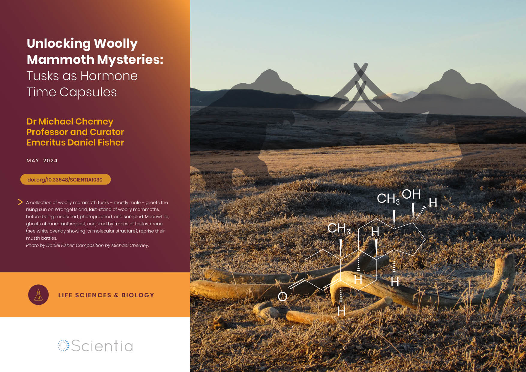
Dr Michael Cherney – Professor Daniel Fisher | Unlocking Woolly Mammoth Mysteries: Tusks as Hormone Time Capsules
The impressive tusks found on proboscideans (the order of mammals that includes elephants, woolly mammoths, and mastodons) are like time capsules, preserving detailed records of their bearers’ lives in the form of growth layers and chemical traces. Frozen in time for thousands of years, these layers can unlock secrets about the lives of long-extinct relatives of modern elephants. Dr Michael Cherney and Professor Daniel Fisher from the University of Michigan used innovative techniques to extract and analyse steroid hormones preserved in woolly mammoth tusks. This ground-breaking work opens new avenues for exploring the biology and behaviour of extinct species.