
Establishing Methods for Medical Imaging and Research: Collaborative Research Centre 1340
As medicine progresses, new techniques are needed to visualise abnormal extracellular structures with greater specificity and resolution. Currently, there is a clear lack of molecular tools to image extracellular structures with the detail needed for early diagnosis of various medical conditions. Based at the Charité – Universitätsmedizin Berlin, the Collaborative Research Centre 1340 represents a large collaboration of researchers from institutions across Berlin, who are working to establish new methods for medical imaging and research at the anatomical and molecular levels.
Developing New Imaging Probes to Better Understand Disease
In medical imaging, techniques such as magnetic resonance imaging use agents that enhance contrast for clearer and more effective images. However, these substances are in most cases non-specific. Disease-specific imaging is needed to improve treatment planning and to improve the assessment of the course of an illness during therapy. However, very few specific agents or probes have been approved for clinical imaging in the last 30 years.
The Matrix in Vision Collaborative Research Centre (CRC) has brought together different researchers from across Berlin. Specialising in various areas of biochemistry, physics and medicine, the researchers focus on the establishment of novel imaging techniques targeting the extracellular matrix (ECM). This is critical due to the role of the ECM in the development and progression of disease, as we will now consider.
The Extracellular Matrix
Most of the recent imaging agents and probes have been components that bind to the cell surface. After the cell surface, the ECM is the next important tissue component that can be targeted by different imaging techniques. The ECM mainly determines the biomechanical properties of tissue and is, therefore, a suitable target for molecular and biophysical imaging approaches.
The ECM functions as a scaffold in which cells of the tissues and organs of the body are embedded. In addition to providing structure, the ECM facilitates interactions between cells including signal transduction between them, cell migration, growth and cell death, as well as immunological functions. It contributes to the regulation of pH as well as hydration.
When tissues are inflamed, injured or invaded by tumours, the ECM is involved in an adaptive response, remodelling itself in defence. This remodelling involves changes in the ECM’s biochemical composition and physiomechanical properties (i.e., the rigidity or elasticity of the scaffold). Due to the changes the ECM undergoes in diseased tissue, it is of interest as a target for in vivo (in the body) imaging approaches for the detection, characterisation and monitoring of disease. The CRC focuses on novel imaging techniques for the characterisation of the ECM.
The main structural components of the ECM include collagen, elastin and glycoproteins/proteoglycans. Collagen is a component of the skin, cartilage, tendons, ligaments, and bones and does not stretch much. When damage occurs as a result of inflammation or other trauma, the amount of collagen in the tissue can increase and results in fibrosis and scar formation, leading to a decrease in tissue elasticity. The protein elastin is a main component of the blood vessel walls. Tissue inflammation can lead to changes in the content of elastin in the ECM.
Proteoglycans (PGs) are a major component of the ECM. PGs consist of a core protein associated with carbohydrate groups consisting of glycosaminoglycans (GAGs). GAGs have a strong negative charge, which allows them to bind water and exert an influence on tissue properties. In different pathological processes, including inflammation and tumour invasion, the amount of one or more of the different types of GAGs in the ECM can be increased. An important characteristic of the GAGs is their ability to form complexes with positively charged molecules.
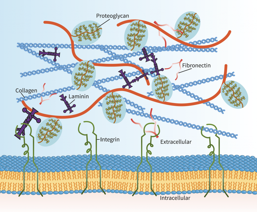
Illustration of the extracellular matrix – after the cell, this is the next important tissue component that can be targeted by different imaging techniques.
Goals of the CRC
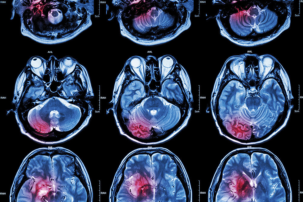
Inflammation and fibrosis occur in different diseases, such as (artery plaques), heart disease, multiple sclerosis, and inflammatory conditions of the intestine and liver. In all these diseases, changes in the ECM occur. The long-term goal for the CRC is the development of new imaging techniques for the characterisation of the ECM.
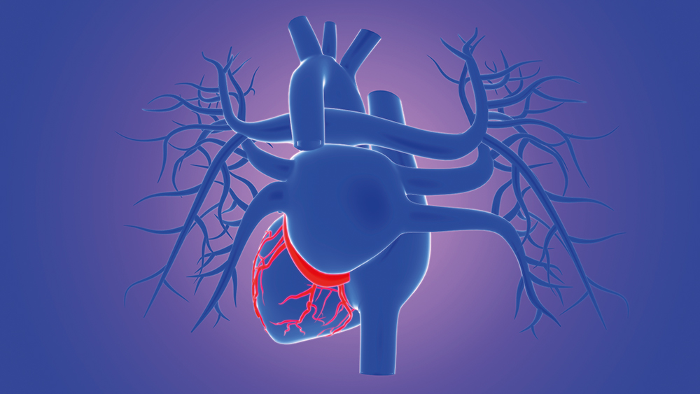
In heart disease, diabetes and hypertension, damage to the heart leads to remodelling of the ECM, which includes the increased formation of PGs and GAGs in order to regulate inflammation, fibrosis, and new blood vessel formation. An increase in collagen affects the biomechanical properties of the heart muscle, leading to a more rigid tissue.
The ECM also plays a role in the central nervous system, maintaining the structural integrity of the tissue through its interactions with various nerve and inflammatory cells.
In Crohn’s disease, GAGs and collagen can increase in inflamed sections of the bowel wall, leading to fibrosis accompanied by fibrosis of the bowel wall.
In the development of liver fibrosis and cirrhosis of the liver, the composition of the ECM becomes impaired at an early stage due to a change in the synthesis and degradation of ECM components. The collagen content rises with the degree of fibrosis, resulting in an increase in tissue rigidity. There is also a distinct increase in a number of types of GAGs found in the fibrotic liver.
The researchers at the CRC 1340 include experts in the fields of diagnostic imaging, medical technology, nanotechnology, cardiovascular disease, neurology, and internal medicine. The group will investigate new imaging approaches for the imaging-based characterisation of the ECM.
Reference
https://doi.org/10.33548/SCIENTIA638
Meet the researcher

Collaborative Research Centre 1340
Charité – Universitätsmedizin Berlin
Berlin
Germany
The Matrix in Vision Collaborative Research Center (CRC 1340) is a branch of the German Research Foundation consisting of a collaboration of 27 principal researchers from the Technical University of Berlin, Max-Planck Institute of Colloids and Interfaces, the National Metrology Institute of Germany, and the National Institute for Material Research and Testing. With a combined expertise encompassing many fields across physics, cell biology and biochemistry, the researchers share the common goal of investigating how different extracellular matrix components can be targeted for in vivo imaging using inflammation as a disease model. By experimenting with in vitro and in vivo model systems, combining molecular methods in radiology with new insights into how mechanical tissue parameters affect the development of disease, the CRC will investigate new molecular imaging probes and imaging approaches for a variety of clinically relevant inflammatory diseases.
SPOKESPERSON
Professor Bernd Hamm
E: bernd.hamm@charite.de
W: https://sfb1340.charite.de/en/
FUNDING
German Research Foundation (DFG)
FURTHER READING
Y Kobayashi, R Hauptmann, H Kratz, et al, Europium doping of superparamagnetic iron oxide nanoparticles enables their detection by fluorescence microscopy and for quantitative analytics, Technology and Health Care, 2017, 25, 457–470.
RL Lindquist, S Papazoglou, C Scharlach, et al, Imaging of magnetic microfield distortions allows sensitive single-cell detection, Molecular Imaging, 2013, 12, 83–89.
I Sack I, K Jöhrens, J Würfel, J Braun J, Structure-sensitive elastography: On the viscoelastic powerlaw behavior of in vivo human tissue in health and disease, Soft Matter, 2013, 9, 5672–5680
MR Makowski, G Varma, AJ Wiethoff, et al, Noninvasive assessment of atherosclerotic plaque progression in apoe-/- mice using susceptibility gradient mapping, Circulation: Cardiovascular Imaging, 2011, 4, 295–303.
E Tysiak, P Asbach, O Aktas, et al, Beyond blood brain barrier breakdown – in vivo detection of occult neuroinflammatory foci by magnetic nanoparticles in high field MRI, Journal of Neuroinflammation, 2009, 6, 20.
![]()
Want to republish our articles?
We encourage all formats of sharing and republishing of our articles. Whether you want to host on your website, publication or blog, we welcome this. Find out more
Creative Commons Licence
(CC BY 4.0)
This work is licensed under a Creative Commons Attribution 4.0 International License. 
What does this mean?
Share: You can copy and redistribute the material in any medium or format
Adapt: You can change, and build upon the material for any purpose, even commercially.
Credit: You must give appropriate credit, provide a link to the license, and indicate if changes were made.
More articles you may like
Probing Electron Dynamics in the Ultrafast Regime
In the atoms that make up the matter around us, negatively charged particles called electrons have properties such as spin and orbital angular momentum. Researchers at Martin Luther University Halle-Wittenberg have developed a theoretical framework which allows them to simulate the dynamics of the spin and orbital angular momentum of electrons in materials when probed with an ultrafast laser pulse. Using this framework, they are able to simulate different materials and improve our understanding of dynamics on an atomic scale.

Seeing Beneath the Surface: Exploring Deltaic Reservoirs with Augmented Reality
In the Aínsa Basin of the Spanish Pyrenees, the Mondot-1 well was drilled, cored, and fully logged to capture a detailed record of a long-buried ancient river delta system. Dr. John D. Marshall, Dr. Jürgen Grötsch, and Dr. Michael C. Pöppelreiter with co-workers at Shell International used this core to trace how sediments once flowed across the landscape, and were deposited under shifting tectonic conditions. The team employed augmented reality and interactive virtual displays; these innovative tools offer new ways to explore subsurface depositional systems, and are particularly useful in locations where physical access to the core is difficult, or no longer possible.
Dr Jim Wu | Ziresovir Offers New Hope for Treating Respiratory Syncytial Virus Infections
Respiratory syncytial virus (RSV) causes respiratory tract infections in children and adults. While for many patients the outcomes of infection are mild, for others, infection can prove fatal, and there is a lack of effective treatments. Dr Jim Wu from the Shanghai Ark Biopharmaceutical Company in China carries out his vital research to develop new, safe, and effective treatments to tackle this killer.
Dr Sandra Grumelli | The Importance of the Choline in Chronic Lung Infections
People with chronic lung conditions like COPD and cystic fibrosis are vulnerable to lung infections caused by the bacterium Pseudomonas aeruginosa. These infections are often difficult to treat and can cause sudden worsening of symptoms, known as flare-ups or acute exacerbations. While we know P. aeruginosa triggers inflammation and damage in the lungs, much less is understood about how exactly it causes these flare-ups, or how it survives in such a harsh environment. Dr Sandra Grumelli from the Center of Investigations of Respiratory Diseases in Argentina, has explored the role of a common molecule called choline which is released during infection. Using a combination of mouse models and laboratory experiments, she has discovered that choline not only makes breathing harder, it also helps P. aeruginosa adapt to and persist in the lungs. Her research opens up new possibilities for tackling chronic infections by targeting the bacteria’s energy use and the way it responds to its environment.