
Professor Deborah Ferrington – Uncovering The Mysteries Of Agerelated Macular Degeneration
Age-related macular degeneration is the leading cause of blindness amongst the elderly population in the developed world. Professor Deborah Ferrington and her colleagues at the University of Minnesota are carrying out ground-breaking research on the cellular pathways underlying the pathology of this life altering disease.

The Fight Against Blindness
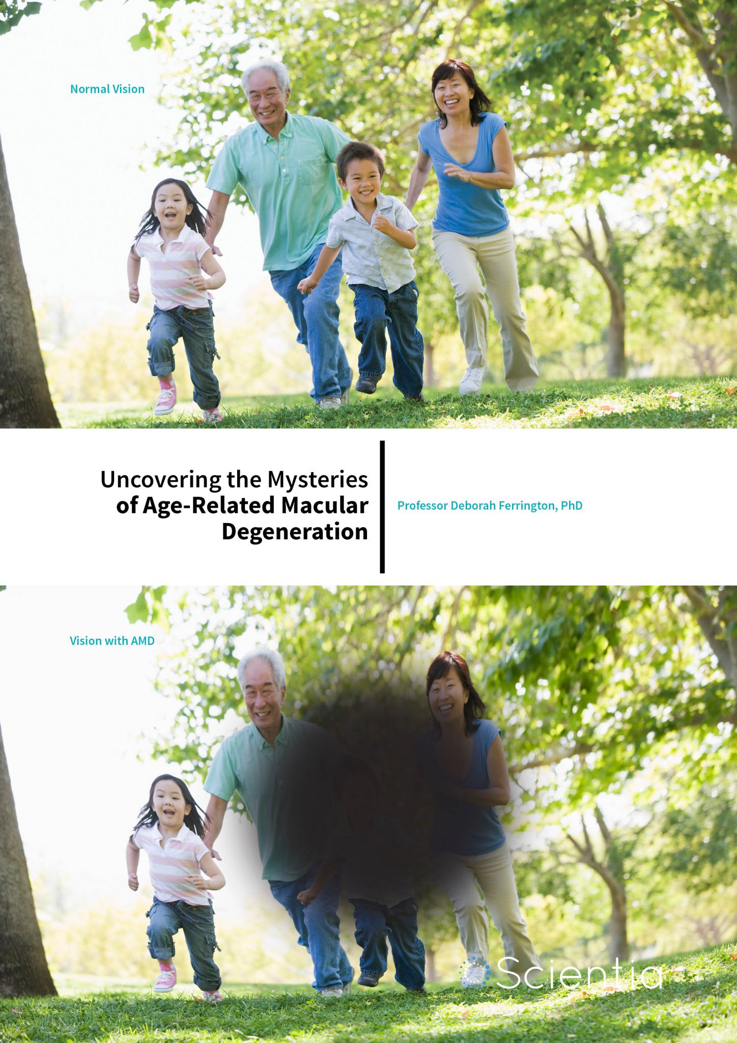
Age-related macular degeneration (AMD) is a life changing disease which robs people of central vision, limiting their ability to read, drive and even recognise faces. AMD affects over 10 million individuals in the United States and this number is expected to double by 2050 due to the Western world’s rapid increase in the ageing population. With limited treatment options available, there is an urgent need to develop new methods for prevention and cure.
AMD leads to vision loss through the destruction of the macula, an oval shaped pigmented area at the centre of the retina. The macula contains a dense concentration of cone cells (which supply high acuity and colour vision). As these cones degenerate, central vision is lost. There are a number of factors which contribute to cone loss, including age, genetic profile and environmental insults such as smoking.
There are two primary forms of AMD. ‘Wet’ AMD manifests as the abnormal growth of blood vessels into the retina. There are successful treatment options available for individuals with this form of the disease, however only 10% of patients present with ‘wet’ AMD. The other type is ‘dry’ or atrophic AMD, which involves changes in and the eventual loss of cells from the retinal pigment epithelium (RPE). The RPE is a monolayer of cells located beneath the neural retina. The RPE maintains retinal health and homeostasis by supporting the function of rod and cone photoreceptors (structures in the eye that respond to light). AMD leads to the formation of lipoproteinaceous deposits (known as drusen) that form between the RPE and choroid (the pigmented vascular area beneath the RPE). As the disease advances, drusen increases in quantity. Changes to the RPE become evident in the early stages of AMD and because these cells are postmitotic, which means they are unable to divide, they cannot be replaced if damaged or lost. Without support from the RPE, photoreceptors die and vision loss occurs. There is currently no effective treatment for ‘dry’ AMD. This is where the work of Professor Deborah Ferrington comes in. Her laboratory is focused on defining molecular changes that occur
in the retina with AMD. ‘The ultimate goal of my research is to identify therapeutic targets for treating AMD, which requires a thorough understanding of the disease mechanism,’ she explains. Defining the molecular mechanism of AMD is no small task. Because the anatomical structure of the retina is unique to primates, no animal models are available which can faithfully replicate the retinal conditions associated with AMD. Therefore, Professor Ferrington and her team are using human donor tissues to study the disease. They aim to investigate AMD pathogenesis through studying changes in protein expression and in the mitochondria at progressive stages of AMD. ‘It was previously thought that one simple mechanism would cause AMD, but it’s not that way at all,’ Professor Ferrington says. ‘Genes implicated in AMD are clustered in several distinct biochemical pathways. You can develop what looks like the same disease many different ways, which is one of the biggest challenges in finding a treatment.’

‘If we can keep the mitochondria healthy, we may slow the progression to blindness… Because mitochondrial damage occurs before vision loss, early intervention would likely protect or rescue RPE mitochondrial function. This is the best place to start aiming therapies.’
Deciphering Damage to Mitochondrial DNA
 Over the last decade, Professor Ferrington and her colleagues have been uncovering evidence that mitochondrial dysfunction is associated with AMD pathogenesis. The mitochondria are organelles found in the cell in which respiration and energy production occur. They also contain a small amount of DNA, separate from the DNA found in the nucleus. Mitochondrial DNA (mtDNA) contains 37 genes which are responsible for coding 13 proteins essential for producing energy and all of the machinery required to make those proteins. The remaining ~1800 proteins that reside in the mitochondria are encoded by nuclear DNA, produced outside of the mitochondria, and then transported into the mitochondria.
So, what evidence is there to suggest that mitochondrial damage may play a role in AMD? Mitochondria are a major source of superoxide anions in the cell. These anions generate highly toxic radicals and hydrogen peroxide that can damage the cell by reacting with proteins, DNA and lipids. This oxidative stress may play an important role in disease progression, as suggested by the increased levels of antioxidant enzymes that occur in response to the oxidative stress and protein adducts (generated from carbohydrate and lipid oxidation) found in AMD donor eyes. mtDNA is more susceptible to damage from oxidation than nuclear DNA and when mtDNA is damaged, this may interfere with production of key proteins involved in energy production.
Based on this evidence, the team analysed the sub-set of proteins present in the mitochondria isolated from the RPE. They found that a significant number of affected proteins were subunits of the mitochondrial ATP synthase complex (which is responsible for energy production). These results suggest specific pathological mechanisms involving altered translation of the ATPase subunits encoded by either nuclear DNA or mtDNA as well as defects in the transport of nuclear encoded proteins into the mitochondria. ‘Mitochondria are a really good target for therapy,’ says Professor Ferrington. ‘We see mitochondrial dysfunction in Parkinson’s and Alzheimer’s diseases, so mitochondria are like an Achilles’ heel for several age-related diseases.’
Normal Ageing or AMD?
A 2010 study provided further evidence that AMD development damages mtDNA inside RPE cells. Professor Ferrington and her team evaluated the extent of mtDNA damage in human RPE for donors of different ages but who did not have AMD to determine the effect of ageing and compared those results with damage in donors with AMD. The purpose was to distinguish damage associated with normal ageing from damage due solely to AMD.
Identifying Targets for Treatments that Stop Vision Loss
 The results showed that mtDNA was limited to the RPE and, as this group had previously shown, lesion frequency increased with disease severity. In contrast, disease stage had no effect on lesions content in the neural retina. ‘That was a complete surprise,’ Professor Ferrington tells us. ‘I had expected that with high damage in RPE the same result would be observed in the neural retina, but that was not so.’ The observation that the retina does not accumulate mtDNA damage with disease progression suggests differences in how AMD manifests in specific tissues. Another important finding was that mtDNA damage was not limited to the macula but was equally abundant in the RPE cells in the peripheral region. This result dispelled a long-held belief that damage only occurs in the macula. Finally, measures of mtDNA damage in small segments of the mitochondrial genome shows that over half of the mitochondrial genome is not significantly damaged in AMD, refining the results of the team’s previous reports of genome wide damage to more discrete regions.
These results are important clinically relevant findings – there is now a scientific basis for targeting RPE mitochondria as a treatment strategy. Because this damage occurs before vision loss, early intervention could prevent or at least slow down progression to blindness.
Genetic Risk for Increased mtDNA Damage
Previous genetic analysis of AMD had identified a number of high risk loci associated with the disease. The genes at these loci belong to diverse pathways, suggesting different pathogenic mechanisms lead to the clinical manifestation of the disease and implies that therapies targeting a single pathway will not be effective for all AMD patients. This idea provided the rationale for a 2016 study aimed to determine if individuals with a specific genetic background were at greater risk for mtDNA damage.
What Comes Next?
Meet the researcher

Professor Deborah Ferrington, PhD
Department of Ophthalmology and Visual Neurosciences
University of Minnesota, Twin Cities, USA
CONTACT
E: ferri013@umn.edu
T: (+1) 612 624 8267
W: http://www.ophthalmology.umn.edu/bio/ophthalmologydepartment-
facul/deborah-ferrington
KEY COLLABORATORS
Dale Gregerson, PhD, University of Minnesota
Sandra Montezuma, MD, University of Minnesota
Erik van Kuijk, MD, PhD, University of Minnesota
James Dutton, PhD, University of Minnesota
Anand Swaroop, PhD, National Eye Institute
Rinki Ratnapriya, PhD, National Eye Institute
M. Christine Kenney, MD, PhD, University of California at Irvine
Robert Mullins, PhD, University of Iowa
FUNDING
Minnesota Regenerative Medicine
National Institutes of Health – National Eye Institute, National Institute of Aging
American Federation for Aging
American Health Assistance Foundation
Arnold and Mabel Beckman Foundation
Foundation Fighting Blindness
Elaine and Robert Larson Endowed Vision Research Chair
Minnesota Lions Vision Foundation
Helen Lindsay Family Foundation
Anonymous Donor for AMD Research
REFERENCES
DA Ferrington, RJ Kapphahn, MM Leary, SR Atilano, MR Terluk, P Karunadharma, G Kuei-Jie Chen, R Ratnapriya, A Swaroop, SR Montezuma and MC Kenney, Increased retinal mtDNA damage in the CFH variant associated with age-related macular degeneration, Experimental Eye Research, 2016, 145, 269–277.
MR Terluk, RJ Kapphahn, LM Soukup, H Gong, C Gallardo, SR Montezuma and DA Ferrington, Investigating Mitochondria as a Target for Treating Age-Related Macular Degeneration, The Journal of Neuroscience, 2015, 35, 7304–7311.
PP Karunadharma, CL Nordgaard, TW Olsen and DA Ferrington, Mitochondrial DNA Damage as a Potential Mechanism for Age-Related Macular Degeneration, Investigative Ophthalmology & Visual Science, 2010, 51, 5470–5479.
CL Nordgaard, PP Karunadharma, X Feng, TW Olsen and DA Ferrington, Mitochondrial Proteomics of the Retinal Pigment Epithelium at Progressive Stages of Age-Related Macular Degeneration, Investigative Ophthalmology & Visual Science, 2008, 49, 2848–2855.

Download