
Professor Gordon Carmichael – Unravelling the Biology of snoRNAs Implicated in Prader-Willi Syndrome
Prader-Willi Syndrome is a rare genetic neurodevelopmental disorder that gives rise to a vast array of symptoms which affect the individual from birth. There is currently no cure for Prader-Willi Syndrome. Professor Gordon Carmichael and his team from the Department of Genetics and Genome Sciences at the University of Connecticut Health Centre, USA, believe it is crucial to understand the affected chromosome 15 region to unravel the pathogenesis of Prader-Willi Syndrome and his team are making significant strides towards achieving this goal.
Prader-Willi Syndrome
Prader-Willi Syndrome (PWS) is caused by the loss of function of a specific area of the paternal chromosome 15 – specifically, q11-q13 – due to deletions or mutations of this region. PWS is diagnosed at birth when symptoms include hypotonia (low muscle tone), feeding difficulty, poor growth and delayed development. In childhood, patients develop hypogonadism (a decreased production of sex hormones by the ovaries or testes) and an extreme appetite (hyperphagia) leading to overeating and obesity. Other symptoms include learning disabilities, behavioural problems, sleep abnormalities and underdeveloped genitals.
Adults with PWS are unable to live fully independent lives and the development of obesity due to excessive eating means sufferers are more likely to develop type 2 diabetes, heart failure and respiratory problems. There is no cure for PWS and current treatments are designed only to manage symptoms such as overeating and behavioural problems. Life-expectancy is considered to be around 29 years.
PWS is a complex disorder, the intricacies of which are still unclear. However, understanding of the molecular mechanisms that underlie PWS can potentially change the management and treatment of the disorder and improve the quality of life of sufferers. Professor Gordon Carmichael and his team at the Department of Genetics and Genome Sciences, University of Connecticut Health Centre, are leading the way in characterising the genetic and molecular defects that underlie PWS to help elucidate the mechanisms of this intriguing disease.

The Discovery of New RNAs in PWS Pathogenesis
Professor Carmichael explains that the 15q11-q13 critical region is ‘extremely complexed, generating a number of both coding and noncoding RNAs.’ Among the important non-coding RNAs expressed from this region are snoRNAs (small nucleolar RNAs), which are conserved nuclear RNAs between 70–200 nucleotides long and are believed to function in the modification of small nuclear RNAs (snRNAs) or ribosomal RNAs (rRNAs), or may be involved in rRNA processing during ribosome biogenesis. snoRNAs are encoded in the introns of protein-coding genes and several hundred snoRNAs are known to exist.
snoRNAs have been reported to be involved in the function and development of the brain. Moreover, those in the chr15q11-q13 region are not expressed from the paternal chromosome of PWS patients or PWS mouse models, indicating a key role of snoRNAs in PWS. While most snoRNAs are excised from debranched introns by a process called exonucleolytic trimming, and then form complexes with specific protein components to form snoRNPs (ribonucleoprotein complexes) and carry out their functions, in a 2012 research paper, Dr Carmichael’s team provided an account of how snoRNAs can also lead to the processing of a new class of long non-coding RNAs.
The many snoRNAs can be divided into two main classes: box C/D snoRNAs and box H/ACA snoRNAs. The box C/D snoRNAs guide 2’-O-methylation and box H/ACA snoRNAs guide pseudouridine modifications. Many researchers agree that snoRNAs are key to PWS. However, the function of the snoRNAs in the PWS critical region and on the function of neurons in the brain is unknown.
Within the 15q11-q13 critical region there lies a cluster of about 30 C/D box snoRNAs – this cluster is called SNORD116. SNORD116 is highly implicated in PWS as all reported deletions and mutations in PWS affect this cluster. ‘The problem with the SNORD116 snoRNAs’ Professor Carmichael explains, ‘is that they are orphans’ since they do not have complementarity to rRNA or other known RNAs and thus their targets are unknown. Therefore, it is important to identify the targets of the SNORD116 snoRNAs and their functions to understand their crucial role in PWS.
‘Finding sites of modification will be exciting, but the real challenge that lies ahead is to then figure out how such modifications affect RNA function and are connected to PWS pathology.’

Developing a 2’-OMe Detection Method to Find Sites Targeted by SNORD116 snoRNAs
The SNORD116 cluster of box C/D snoRNAs is highly expressed in the brain and the importance of these types of snoRNAs in PWS pathology is striking. The team already knew that box C/D snoRNAs modify RNAs by 2’-OMe, but strategies to map sites of 2’-OMe on RNA molecules were inefficient.
In 2017, Professor Carmichael and his team developed a new method, RibOxi-seq, to map sites of 2‘-OMe to better understand the targets of the SNORD116 snoRNAs. Previous methods lacked specificity and relied on negative rather than positive signals. Using next-generation sequencing, the team developed a highly sensitive and accurate method to detect methylation relying on positive signals.
The basic structure of RNA is a five-carbon sugar attached to one of four nitrogenous bases; A, G, U or C, and this unit of sugar and base is called a nucleotide. An RNA primary structure is made up of a single-stranded chain of nucleotides linked together by phosphodiester bonds. The fifth carbon of the sugar carries an unbound phosphate group and is thus called the 5’end. As the last sugar at the other end of the nucleotide chain has a free hydroxyl (-OH) group at the third carbon, this end is called the 3’ end, hence RNA molecules have a 5’ and 3’ end.
In the RibOxi-seq method, Professor Carmichael’s team reported that RNA fragments are generated using random 3’-ends, followed by periodate oxidation (a reaction to split bonds between carbons) of all molecules terminating in the 2’,3’-OH groups. In this way, only RNA that harbours 2’-OMe groups at their 3’-ends are intact to be sequenced. Once sequenced, the RNA is aligned to a reference genome in the UCSC Genome Browser and the data is analysed for enrichment. The team tested the RibOxi-seq method to analyse RNA from the human teratoma-derived PA-1 cell line, a cell line that is known to highly express SNORD116 snoRNAs.
As a result of these experiments, not only was the team able to detect known 2’-OMe sites in model ovarian carcinoma PA-1 cells but also to identify new sites. RibOxi-seq was confirmed to be a highly sensitive and accurate method to detect the modification of RNA by 2’-OMe. The researchers further discussed that there are, however, still some limitations to the method, such as the need for microgram levels of starting material. Also, RNAs that are shorter than 100 base pairs (bp) are more difficult to study, as the fragments needing to be generated would be very small. Professor Carmichael shares with us that it’s also ‘much harder to map sites in mRNA which is less abundant and very sequence diverse. But we’re getting there.’ Having initially studied 2’-OMe in PA-1 cells, the team’s next goal is to extend these studies to PWS.

Harnessing Neurons as a Model to Study PWS
SNORD116 is expressed at high levels in human stem cells and neurons which make them both ideal cells to study the critical PWS region. Using a human stem cell line, Dr Stormy Chamberlain, one of Dr Carmichael’s colleagues, utilised a technology called CRISPR/Cas9 to modify one line of an isogenic pair of stem cells by deleting the paternal PWS critical region. These modified stem cells lacking the critical region, along with their isogenic partners with the critical region still intact, are differentiated into neurons. The team have been performing mRNA-sequencing of extracted RNA from these neurons which will help to identify the differential expression of RNAs between cells with and without SNORD116. Alongside these experiments, the team is also using their recently developed RibOxi-seq method to map 2’-OMe sites and identify SNORD116 snoRNA targets in these cells.
These experiments aim to gain greater insight into the SNORD116 targets and their role in the pathogenesis of PWS. Professor Carmichael believes ‘finding sites of modification will be exciting, but the real challenge that lies ahead is to then figure out how such modifications affect RNA function and are connected to PWS pathology.’

Implications for Other Diseases
As very little is known about the effects of 2’-OMe modification in mRNA, Professor Carmichael believes this work will also lead to a new understanding of snoRNA function in biology. Furthermore, as hundreds of orphan snoRNAs are known to be expressed in the genome, some snoRNAs have also been linked to other diseases and disorders such as cancer. The wider implications of Professor Carmichael’s research are the broader understanding of the biology of snoRNAs and 2’-OMe which may lead to a better understanding of other diseases and potentially the development of new treatments in the future.
Reference
https://doi.org/10.33548/SCIENTIA547
Meet the researcher

Professor Gordon Carmichael
Department of Genetics and Genome Sciences
University of Connecticut Health Centre
Farmington, Connecticut
USA
Professor Gordon Carmichael is currently a Professor of Genetics and Genome Sciences at the University of Connecticut Health Centre. Professor Carmichael obtained his PhD from Harvard University in 1975 and has a longstanding interest in the molecular mechanisms controlling the function and expression of RNA molecules. At present, his laboratory focuses on the functions of a new class of long noncoding RNA molecules implicated in the pathogenesis of Prader-Willi Syndrome and on small noncoding RNAs expressed from the same genomic locus. During his career, Professor Carmichael was one of the first to use RNA affinity chromatography to purify proteins. He also developed a now widely-used method for RNA gel electrophoresis, and was amongst the first to use synthetic DNA oligonucleotides to produce mutations in viral genes. In addition to these and other achievements, Professor Carmichael also serves as an Editorial Board Member of a number of journals and has been the recipient of numerous honours and awards, representing his outstanding reputation in his field.
CONTACT
E: carmichael@uchc.edu
W: https://facultydirectory.uchc.edu/profile?profileId=3078
KEY COLLABORATORS
Stormy J. Chamberlain, UConn Health
Justin L. Cotney, UConn Health
FUNDING
Foundation for Prader-Willi Research
Grant RO1 HD099975 from the National Institutes of Health
FURTHER READING
Y Zhu, SP Pirnie, GG Carmichael, High-throughput and site-specific identification of 2′-O-methylation sites using ribose oxidation sequencing (RibOxi-seq), RNA, 2017, 23(8), 1303–1314.
QF Yin, L Yang, Y Zhang, et al, Long noncoding RNAs with snoRNA ends, Molecular Cell, 2012, 48(2), 219–30.
![]()
Want to republish our articles?
We encourage all formats of sharing and republishing of our articles. Whether you want to host on your website, publication or blog, we welcome this. Find out more
Creative Commons Licence
(CC BY 4.0)
This work is licensed under a Creative Commons Attribution 4.0 International License. 
What does this mean?
Share: You can copy and redistribute the material in any medium or format
Adapt: You can change, and build upon the material for any purpose, even commercially.
Credit: You must give appropriate credit, provide a link to the license, and indicate if changes were made.
More articles you may like

Dr Lifei Wang | Can Species Distribution Models Inform Us About Future Ecosystems?
The world is buzzing with news about how human activities and climate shifts are reshaping our ecosystems. Have you ever wondered how life will adapt to this rapidly changing world? Ecologists might be able to predict how different species will live in future using computer simulations. Dr Lifei Wang at the University of Toronto Scarborough investigates how different stimulations work under varying conditions to provide new insights into what may lie ahead.
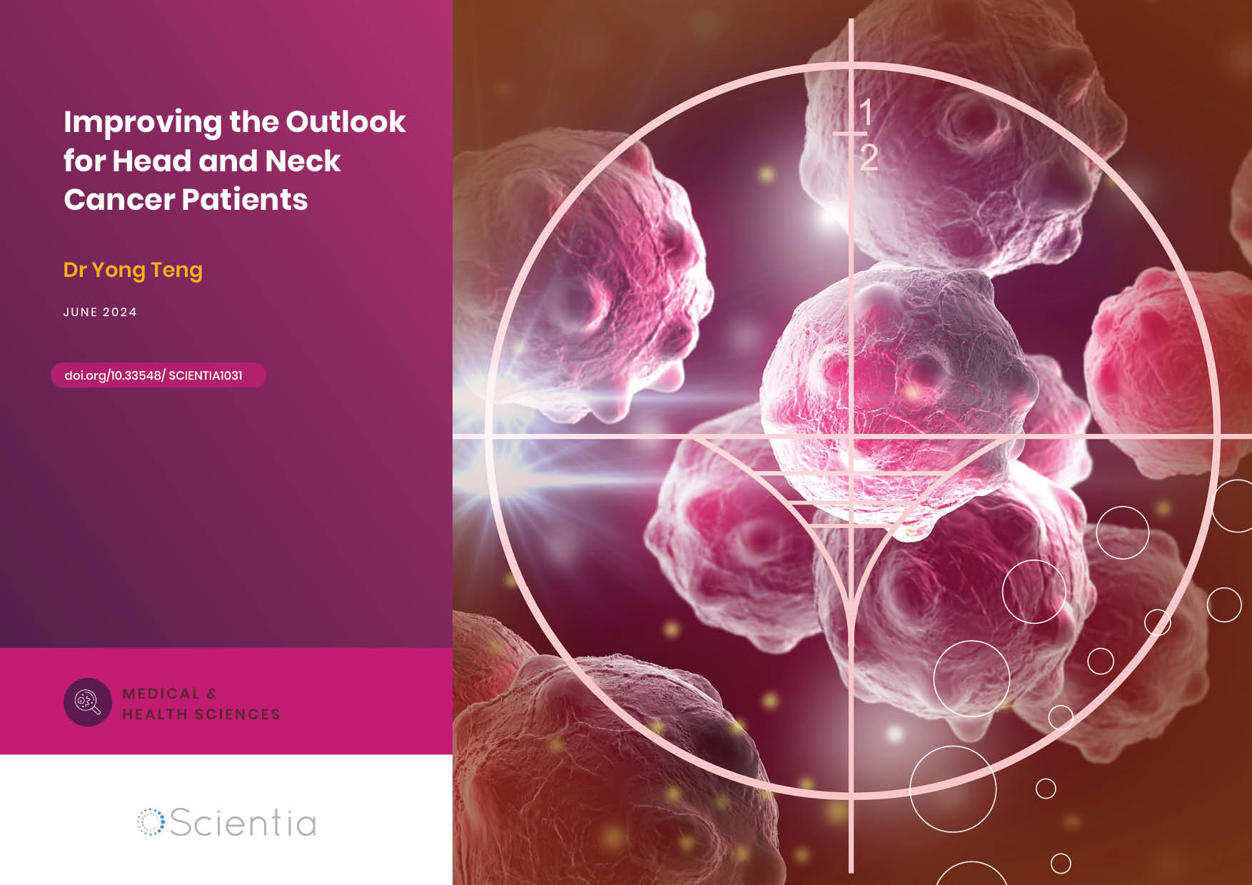
Dr Yong Teng | Improving the Outlook for Head and Neck Cancer Patients
Dr Yong Teng at the Emory University School of Medicine is working with colleagues to overcome the high mortality of individuals diagnosed with cancers affecting the head and neck. One of his approaches is based on understanding the particular mechanisms of the ATAD3A gene, which new insights suggest are closely related to cancers affecting the head and neck.
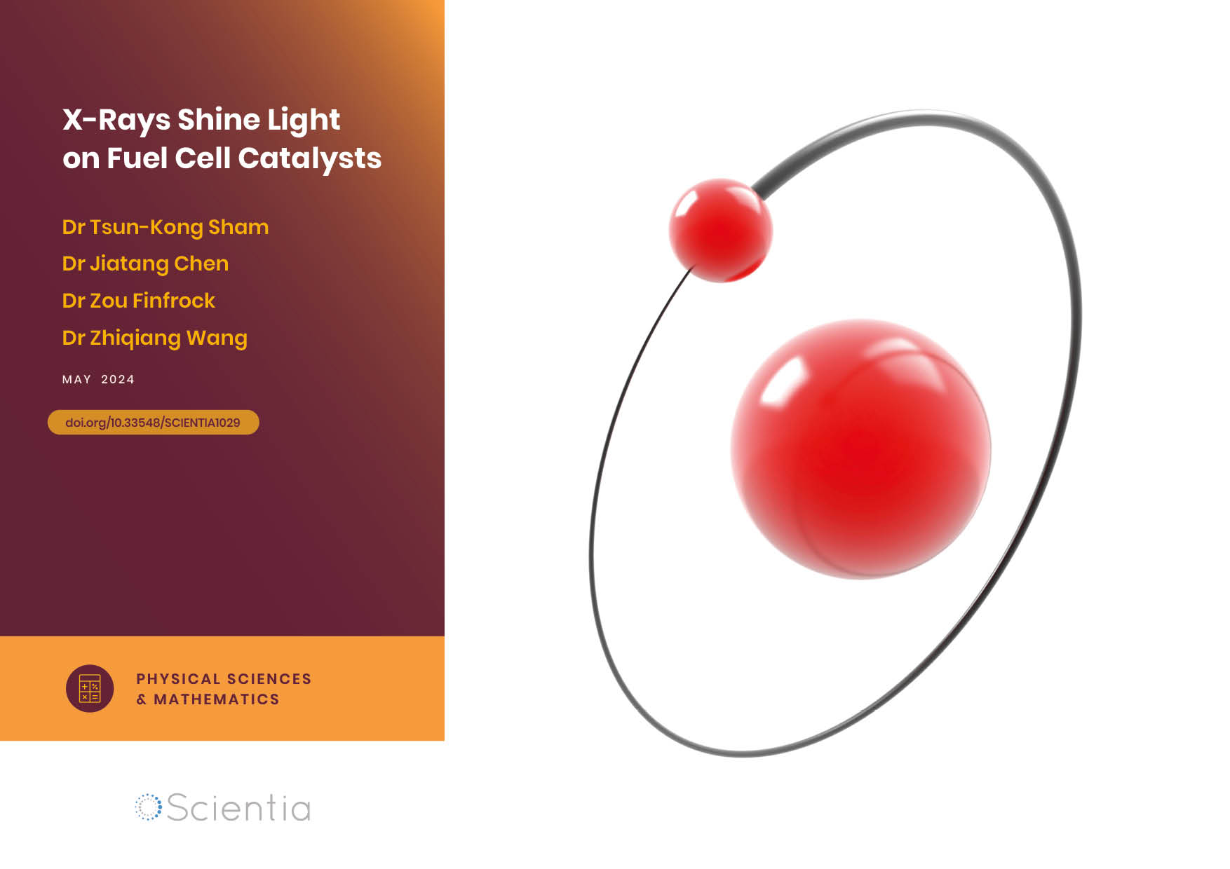
Dr Tsun-Kong Sham – Dr Jiatang Chen – Dr Zou Finfrock – Dr Zhiqiang Wang | X-Rays Shine Light on Fuel Cell Catalysts
Understanding the electronic behaviour of fuel cell catalysts can be difficult using standard experimental techniques, although this knowledge is critical to their fine-tuning and optimisation. Dr Jiatang Chen at the University of Western Ontario works with colleagues to use the cutting-edge valence-to-core X-ray emission spectroscopy method to determine the precise electronic effects of altering the amounts of platinum and nickel in platinum-nickel catalysts used in fuel cells. Their research demonstrates the potential application of this technique to analysing battery materials, catalysts, and even cancer drug molecules.
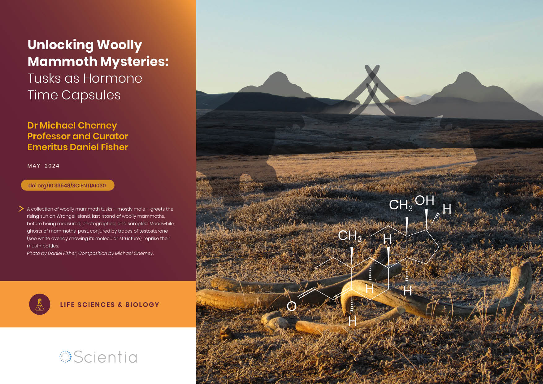
Dr Michael Cherney – Professor Daniel Fisher | Unlocking Woolly Mammoth Mysteries: Tusks as Hormone Time Capsules
The impressive tusks found on proboscideans (the order of mammals that includes elephants, woolly mammoths, and mastodons) are like time capsules, preserving detailed records of their bearers’ lives in the form of growth layers and chemical traces. Frozen in time for thousands of years, these layers can unlock secrets about the lives of long-extinct relatives of modern elephants. Dr Michael Cherney and Professor Daniel Fisher from the University of Michigan used innovative techniques to extract and analyse steroid hormones preserved in woolly mammoth tusks. This ground-breaking work opens new avenues for exploring the biology and behaviour of extinct species.