
Professor Irene Díaz-Moreno – Professor Miguel A. De la Rosa | The Diverse Interactome of Cytochrome c: Beyond Respiration
All living things are comprised of cells, and to function, most of them use oxygen to break down food molecules to obtain chemical energy, a process known as cell respiration. Critical to this is the macromolecule cytochrome c, but this redox haemoprotein also boasts a diverse set of functions beyond respiration. Professor Irene Díaz-Moreno and Professor Miguel A. De la Rosa, both leading members of cicCartuja’s Biointeractomics Research Group at the University of Seville, are using cutting-edge investigational tools to study the full ‘interactome’ of this multifunctional molecule.
Cytochrome c: A Multifunctional Molecule
All complex organisations need communication – whether human societies or living cells. Cells are the building blocks of all living things, and have networks of biomolecules that transmit ‘messages’ necessary for proper functioning. Biointeractomics is a fusion science that aims to unveil the complete set of these biomolecular networks, borrowing from biology, informatics, and engineering disciplines. The Biointeractomics Research Group from the University of Seville, led by Professor Irene Díaz-Moreno and Professor Miguel A. De la Rosa, is at the forefront of this exciting field.
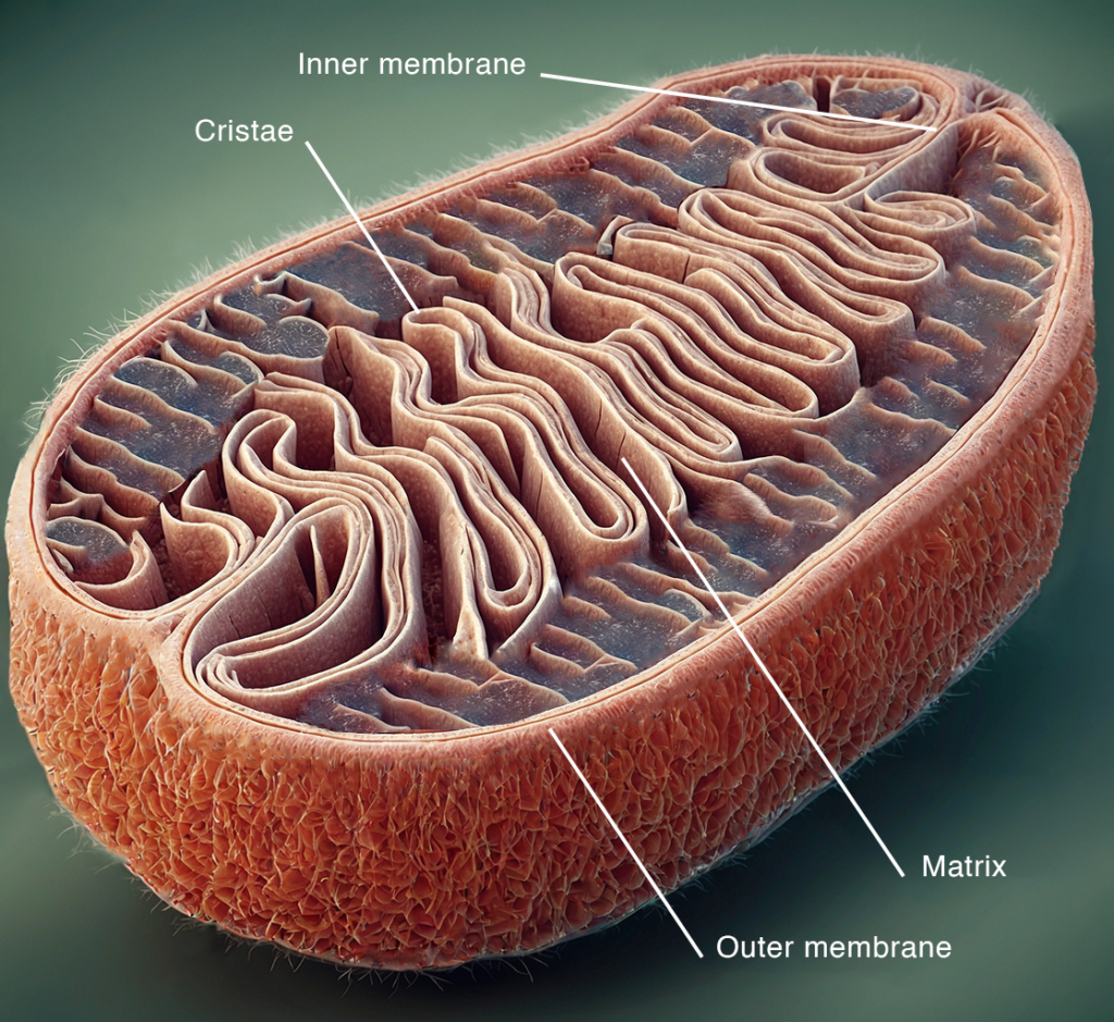
The macromolecule cytochrome c (Cc) is a redox haemprotein usually found in the mitochondrial intermembrane space. It plays a critical role in cell respiration, the process through which organisms use oxygen to break down food molecules to obtain chemical energy to support cell functions.
However, Cc’s activities go beyond respiration. Cc localises in other subcellular structures known as organelles, and has a diverse range of functions, including regulating cell immolation (apoptosis), cell signalling, and scavenging reactive oxygen species (ROS). Professor Díaz-Moreno and Professor De la Rosa’s research group deploys various biochemical, biophysical, and computational tools to study the Cc ‘interactome’, which refers to its full set of interactions.
Probing into Cytochrome c’s Role in Mitochondrial Electron Transfer
The mitochondria, where respiration occurs, are the cell’s ‘powerplants’, supplying energy in the form of adenosine triphosphate (ATP) for vital functions. The electron transport chain (ETC) is the final step of respiration, taking place in the intermembrane mitochondrial space. NADH and FADH2 – produced in the preceding steps of glycolysis, pyruvate oxidation, and the citric acid cycle – supply the electrons for the ETC. Electron transfer (ET) occurs across the integral membrane protein complexes, complex I (NADH coenzyme Q reductase), complex II (succinate dehydrogenase), complex III (cytochrome bc1) and complex IV (cytochrome c oxidase), and then to O2, the final electron acceptor. This creates a proton gradient, which drives the formation of ATP by ATP synthase.
Complexes I, II, III and IV are fixed to the inner mitochondrial membrane, so how can electrons pass between them? Unlike electrical circuits, electrons are not transported between redox proteins in metal wires. Instead, they must contend with the lipidic and aqueous environments of the mitochondrial membrane and matrix, respectively, where ubiquinone and Cc come in – these are both small, soluble molecules, making them ideal carriers of electrons between complexes. Electrons are transferred from complex I and II to III by ubiquinone in the membrane, and from III to IV by Cc in the matrix. A high binding turnover of electron carriers, including Cc, is needed to maintain the cell’s energy needs. Cc must, therefore, bind weakly but with high specificity to its redox partners. The research group has elucidated some of the molecular mechanisms behind this.
The research group has focused on electron transport between Complex III and Cc to date. The cytochrome c1 subunit (Cc1) of III binds to Cc, allowing electron transfer. However, they discovered that Cc1 and Cc don’t need to touch for electrons to pass between them. Electrochemical tunnelling spectroscopy (ETS) experiments showed that a current flows between Cc1 and Cc, even when separated by 10 nm. This implies that electron transfer can occur even before a static protein complex is formed. How could this be? Proteins such as Cc1 and Cc have charged surfaces. A layer of water and ions exists at the surface-aqueous solution interface – the Gouy-Chapman layer. The research group concludes that this layer provides a conduit for electrons to transfer long-distance between Cc1 and Cc. They consider this Gouy-Chapman conduit between Cc1 and Cc to be instrumental in sustaining the ETC.
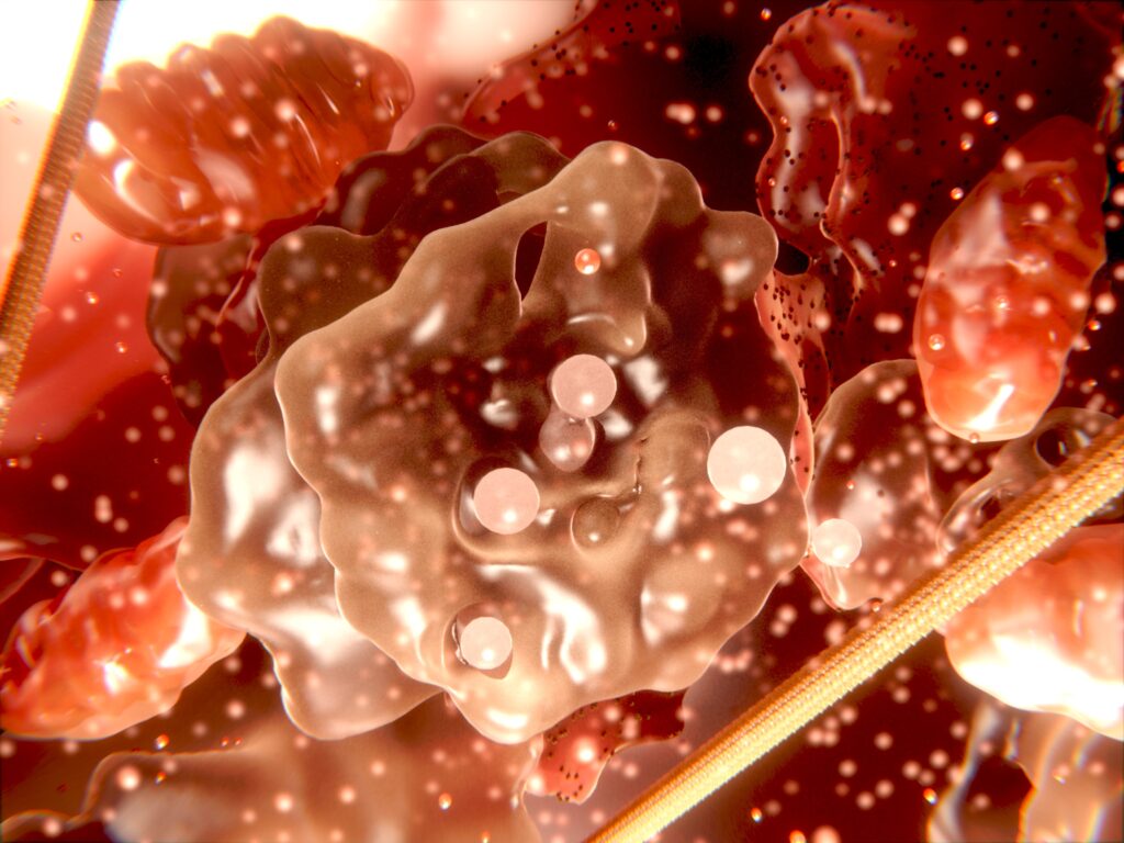
Illustration of cell organelles
Regulating Cytochrome c’s Electron Transfer with Phosphorylation
Sustaining the ETC at the optimum rate is important – but not too fast! Too much electron flux through the ETC can lead to membrane hyperpolarisation, the production of destructive ROS, and the activation of the caspase cascade. This can lead to apoptosis. Luckily, cells have protective mechanisms to regulate this – and Cc has a key role. It is known that Cc phosphorylation downregulates ETC flux, thus protecting cells against apoptotic damage. Phosphorylation mainly happens on serine and threonine residues, but phosphorylation of tyrosine is important in the regulation of signalling and homeostatic functions. In mammals, Cc has six sites for the attachment of phosphate groups – Thr28, Ser47, Tyr48, Thr49, Thr58 and Tyr97. Cc phosphorylation is implicated in various diseases, including ischemia, cancer, and neurodegeneration.
In particular, Tyr48 phosphorylation affects the interactions between Cc and its binding partners, but how this happens was previously unknown. The research group has used a nanoscopic approach to probe into the molecular mechanism of this. It is difficult to isolate in vivo preparations of Tyr48-phosphorylated Cc. So, to mimic the effect of phosphorylation at this position, they deployed a phosphomimetic mutant of Cc, in addition to wild-type Cc. Instead of a Tyr48 residue, they used a variant of Cc that includes a non-canonical amino acid in this position, p-carboxy-methyl-L-phenylalanine (pCMF). pCMF is ideal as it is stable, has a similar volume and charge as phosphotyrosine, and does not affect Cc’s haem moiety.
Wild-type and mutated forms of Cc were expressed in E. coli hosts, purified, and deployed in various experiments. The research group studied Cc–Cc1 binding dynamics using ETS, electrochemical gating, atomic force microscopy, surface plasmon resonance (SPR), single-molecule unbinding experiments, dissociation kinetics studies, and computational simulations. Replacing Tyr48 with pCMF in Cc impaired long-distance electron transfer between Cc1 and Cc. The Y48pCMF (Tyr48-to-pCMF) mutation reduced the ‘decay distance’, at which electrons transferred, by half (3 nm).
Moreover, substituting Tyr48 with pCMF strengthened the interaction between Cc and Cc1, in both single-molecule and bulk-binding experiments. Y48pCMF Cc was shown to have a stronger binding affinity to Cc1 than wild-type Cc binding to Cc1 in SPR studies. Moreover, it was observed that greater force is needed to detach Y48pCMF Cc from Cc1 in single molecule unbinding experiments. The stronger the Cc-Cc1 binding, the lower the turnover – so this stronger binding is thought to slow down Cc turnover. How does this impaired electron transfer and stronger binding translate to respiratory activity? By conducting biochemical assays using mammalian mitochondrial extracts, the group found that Y48pCMF Cc led to a 1.6-fold reduction in complex III activity compared with wild-type Cc.
What relevance do these findings have to the regulation of the ETC? Y48pCMF is a good mimic of phosphorylation at Cc’s Tyr48, so it can be assumed that the findings apply to phosphorylated Tyr48. Both phosphate and pCMF are negatively charged. From molecular dynamics simulations, the research group infers that cations accumulate around the surface of phosphotyrosine, which disrupts the Gouy-Chapman conduit, impeding electron transfer. Furthermore, phospho-Tyr48 Cc is more likely to get stuck at complex III, preventing others from binding and transferring electrons. These results provide mechanistic insights into how cells deploy phosphorylation of Cc to reduce ETC flux, preventing the negative impacts of excess flux, and thus regulating respiration rate.
The Role of Cytochrome c in ARF/p53-mediated DNA Repair
Damage to DNA under stress conditions can lead to loss of function, apoptosis, or even cancer. DNA can be damaged through exposure to mutagenic chemicals, radiation, viral infection, or ROS released in metabolism. Fortunately, cells have several stress responses to limit or mitigate the effects, including the ARF/p53 pathway, activated whenever DNA is damaged. p53 is a tumour suppressor protein that acts through various mechanisms, including activation of DNA repair proteins and cell cycle arrest. The ubiquitin ligase HDM2 (human double minute clone 2) inactivates p53 through ubiquitination and proteosome-dependent degradation. However, this is prevented by ARF (alternative reading frame of the INK4a gene – p14ARF in humans), a protein that inhibits HDM2. Thus, the tumour suppression activity of p53 is negatively and positively regulated by HDM2 and ARF, respectively.
The nucleolus (plural nucleoli) – a membrane-less liquid droplet organelle within the nucleus – is the main ‘traffic controller’ in the cell’s ARF/p53 stress response. In the nucleolus, ARF is stored by being sequestered to nucleophosmin (NPM) protein. Upon DNA damage, ARF is released from the nucleoli, activating p53’s damage control activity. It has long been known that there is crosstalk between the mitochondria and the nucleus, but it was assumed that this was independent of the nucleoli. The group discovered that this crosstalk impacts the nucleoli’s ARF/p53 pathway initiation. What’s more, Cc is at the heart of this. With biophysical and structural analyses, the group probed Cc’s role in the ARF/p53 stress response.
Human cell lines (HeLa cells) were treated with camptothecin (CPT) or doxorubicin (Dox) – compounds that induce single – or double-strand DNA breaks. Immunostaining and proteomic analysis revealed that upon CPT-induced DNA damage, Cc translocates from the mitochondria to the nucleus. Cc was observed to interact with nucleolar NPM in bimolecular fluorescence complementation, coimmunoprecipitation, and proximity ligand assay experiments. In binding studies, they found that Cc competes with p19ARF (the mouse version of ARF) for binding to NPM.
With a suite of structural analyses – including nuclear magnetic resonance, X-ray crystallography, electron microscopy, and computational tools – the group determined the three-dimensional structure of the Cc-NPM binding complex. NPM’s structure comprises an N-terminal structured oligomerisation domain that binds to Cc or p19ARF, a disordered central region that binds to histones, and a C-terminal nucleic acid binding domain. Upon binding to Cc or p19ARF, NPM undergoes liquid-liquid phase separation. In studies involving isolated nucleoli and HeLa nuclei with fluorescently tagged Cc, they observed that Cc displaces p19ARF, which is then released into the cytosol.
These findings are significant in providing a mechanistic explanation for the role of Cc in initiating the DNA damage response (DDR) under stress conditions. Upon DNA damage, Cc translocates from the mitochondria to the nucleus and competitively binds to nucleolar NPM. This releases ARF, which kick starts the ARF/p53 stress response, enabling p53 to activate DNA repair and suppress cancerous tumour formation by arresting the cell cycle.
Future Plans for the Biointeractomics Unit
Through a rich combination of biochemical, biophysical, and computational tools, the Biointeractomics Research Group is discovering that the interactome of Cc is more varied than was thought possible. The observed Cc crosstalk between the mitochondria and nucleus is particularly intriguing. The group plans to delve deeper into Cc’s full range of functions and determine the implications for understanding health and disease.
SHARE
DOWNLOAD E-BOOK

REFERENCE
https://doi.org/10.33548/SCIENTIA1083
MEET THE RESEARCHERS

Professor Irene Díaz-Moreno and Professor Miguel A. De la Rosa
Institute for Chemical Research – Research Centre Isla de la Cartuja (IIQ – cicCartuja), University of Seville – CSIC, Spain
Professor Irene Díaz-Moreno and Professor Miguel A. De la Rosa, both at the University of Seville, are leading members of the Biointeractomics Research Group, Institute for Chemical Research (IIQ), cicCartuja. cicCartuja is a multidisciplinary research centre between the University of Seville, the Spanish National Research Council (CSIC), and the Andalusian Government (Junta de Andalucía). The Biointeractomics Research Group boasts extensive experience in the molecular recognition of metalloproteins, structure-function relationships of signalling proteins, and transient protein-protein and protein-nucleic acid interactions. The Group studies the importance of these interactions in various cellular processes, including the DNA damage response.
Professor Díaz-Moreno joined cicCartuja after a postdoctoral stay in the UK funded by an EMBO fellowship and is the Biointeractomics Group’s team leader. Her research focuses on post-transcriptional and post-translational regulation of DNA damage responses, characterising cytochrome c–histone chaperone interactions. Professor De la Rosa was the Founding Director of the Biointeractomics Unit at the University of Seville’s Institute of Plant Biochemistry and Photosynthesis in 1988, before moving to the IIQ in 2016. His research focuses on structure-activity relationships of biological macromolecules, deploying approaches from various disciplines. In 2014, they founded ManSciTech S.L., a spin-out that deploys metabolomic approaches in commercial applications.
CONTACT
Professor Irene Díaz-Moreno
W: www.iiq.us-csic.es/en/biointeractomics
Professor Miguel A. De la Rosa
E: marosa@us.es
W: www.iiq.us-csic.es/en/biointeractomics
https://www.ae-info.org/ae/Member/De_la_Rosa_Miguel
FURTHER READING
A Lagunas, A Guerra-Castellano, A Nin-Hill, et al., Long distance electron transfer through the aqueous solution between redox partner proteins, Nature Communications, 2018, 9(1), 5157. DOI: https://doi.org/10.1038/s41467-018-07499-x
AMJ Gomila, G Pérez-Mejías, A Nin-Hill, et al., Phosphorylation disrupts long-distance electron transport in cytochrome c, Nature Communications, 2022, 13(1), 7100. DOI: https://doi.org/10.1038/s41467-022-34809-1
K González-Arzola, A Díaz-Quintana, N Bernardo-García, et al., Nucleus-translocated mitochondrial cytochrome c liberates nucleophosmin-sequestered ARF tumor suppressor by changing nucleolar liquid-liquid phase separation, Nature Structural & Molecular Biology, 2022, 29(10), 1024–1036. DOI: https://doi.org/10.1038/s41594-022-00842-3

![]()
![]()
REPUBLISH OUR ARTICLES
We encourage all formats of sharing and republishing of our articles. Whether you want to host on your website, publication or blog, we welcome this. Find out more
Creative Commons Licence (CC BY 4.0)
This work is licensed under a Creative Commons Attribution 4.0 International License. 
What does this mean?
Share: You can copy and redistribute the material in any medium or format
Adapt: You can change, and build upon the material for any purpose, even commercially.
Credit: You must give appropriate credit, provide a link to the license, and indicate if changes were made.
SUBSCRIBE NOW
Follow Us
MORE ARTICLES YOU MAY LIKE
Illuminating Neanderthal Resilience and Adaptability through Cutting-Edge Zooarchaeology
Neanderthals, our closest extinct human relatives, have often been portrayed as brutish and primitive compared to modern humans. But new research is shedding light on their true capabilities. Eboni Westbury from the Australian National University is part of a team investigating how Neanderthals adapted and thrived in challenging Ice Age environments. Their work at the Abric Pizarro rock shelter in Spain reveals new insights into the complex behaviours and survival skills of these ancient people.
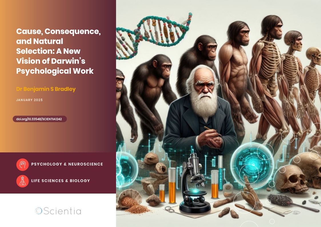
Dr Benjamin Bradley | Cause, Consequence, and Natural Selection: A New Vision of Darwin’s Psychological Work
The theory of evolution by natural selection stands at the heart of modern biology. But what exactly is the causal status of natural selection in evolution? Dr Benjamin Bradley from Charles Sturt University in Australia is challenging long-held assumptions, arguing that Darwin himself saw natural selection as a consequence of other processes, not a cause of evolution in its own right. This crucial distinction opens up new perspectives on how evolution relates to psychology and behaviour.

Professor JoAnne S. Richards | Revealing the Secrets of the Ovary
Professor JoAnne S. Richards conducts her vital research at the Baylor College of Medicine in Houston, Texas, in the USA. For decades she has investigated how certain hormones regulate ovarian function at particular stages of the menstrual cycle, as well as developing new ways to study ovarian cancer. Her work also helps shed new light on common female reproductive health disorders that can affect fertility.
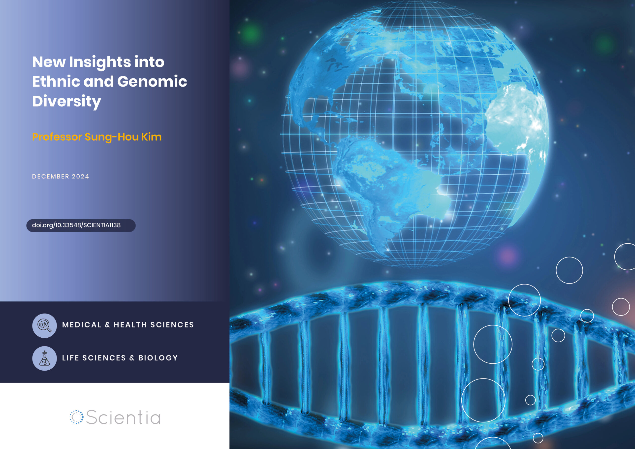
Professor Sung-Hou Kim | New Insights into Ethnic and Genomic Diversity
Does our ethnic diversity translate to genomic diversity? New findings suggest that it might not and point instead to considerable genomic similarities across multiple ethnicities. Professor Sung-Hou Kim at the University of California, Berkeley, and his colleagues classified 164 ethnic groups into 14 genomic clusters spread across various geographical regions. Their findings reveal important new insights into our shared human genetic heritage.